







Preparation And Storage


Recommended Assay Procedures
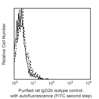
Immunofluorescent Staining and Flow Cytometric Analysis: The purified hTNFR-M1 (Cat. No. 551311) antibody can be used for the immunofluorescent staining (≤ 1 µg antibody/10e6 cells) and flow cytometric analysis of human nucleated cells to measure their expressed levels of surface TNFRII. An appropriate immunoglobulin isotype control is clone R35-38 (Cat. No. 555846). A three-layer staining protocol is recommended for maximizing the detection of TNFRII expressed by cells as detailed in the figure legend.Please note also that as a consequence of in vivo or in vitro activation, cell surface TNFRII can either be shed by cells or transiently expressed at higher levels. As a result, cellular activation can affect the cell's overall expressed level of surface TNFRII.
Product Notices
- Since applications vary, each investigator should titrate the reagent to obtain optimal results.
- Please refer to www.bdbiosciences.com/us/s/resources for technical protocols.
- Caution: Sodium azide yields highly toxic hydrazoic acid under acidic conditions. Dilute azide compounds in running water before discarding to avoid accumulation of potentially explosive deposits in plumbing.
Companion Products

.png?imwidth=320)

The hTNFR-M1 antibody specifically recognizes the extracellular domain of the 75 kDa transmembrane receptor for the human cytokines, tumor necrosis factor (TNF or TNF-α) and lymphotoxin-alpha (LT-α3, aka, lymphotoxin or TNF-β). This receptor is referred to as the p75 or Type II Tumor Necrosis Factor Receptor (TNFRII) [aka, CD120b]. Human TNFRII proteins are expressed by hematopoietic cells including macrophages, neutrophils, lymphocytes, thymocytes and mast cells. TNFRII is expressed by a variety of other cell types including endothelial cells, cardiac myocytes and prostate cells. Naive B cells express very low or undetectable levels of TNFRII whereas mature erythrocytes and platelets are uniformly negative for TNFRII expression. The immunogen used to generate the hTNFR-M1 hybridoma was COS- expressed recombinant human TNFRII.
Development References (11)
-
Aggarwal S, Gollapudi S, Gupta S. Increased TNF-alpha-induced apoptosis in lymphocytes from aged humans: changes in TNF-alpha receptor expression and activation of caspases. J Immunol. 1999; 162(4):2154-2161. (Biology). View Reference
-
Brockhaus M, Schoenfeld HJ, Schlaeger EJ, Hunziker W, Lesslauer W, Loetscher H. Identification of two types of tumor necrosis factor receptors on human cell lines by monoclonal antibodies. Proc Natl Acad Sci U S A. 1990; 87(8):3127-3131. (Biology). View Reference
-
Browning JL, Dougas I, Ngam-ek A, et al. Characterization of surface lymphotoxin forms. Use of specific monoclonal antibodies and soluble receptors.. J Immunol. 1995; 154(1):33-46. (Biology). View Reference
-
Erikstein BK, Smeland EB, Blomhoff HK, et al. Independent regulation of 55-kDa and 75-kDa tumor necrosis factor receptors during activation of human peripheral blood B lymphocytes. Eur J Immunol. 1991; 21(4):1033-1037. (Biology). View Reference
-
Gehr G, Gentz R, Brockhaus M, Loetscher H, Lesslauer W. Both tumor necrosis factor receptor types mediate proliferative signals in human mononuclear cell activation. J Immunol. 1992; 149(3):911-917. (Biology). View Reference
-
Heilig B, Mapara M, Brockhaus M, Krauth K, Dörken B. Two types of TNF receptors are expressed on human normal and malignant B lymphocytes. Clin Immunol Immunopathol. 1991; 61(2):260-267. (Clone-specific: Flow cytometry). View Reference
-
Hohmann HP, Remy R, Brockhaus M, van Loon AP. Two different cell types have different major receptors for human tumor necrosis factor (TNF alpha). J Biol Chem. 1989; 264(25):14927-14934. (Biology). View Reference
-
Munker R, DiPersio J, Koeffler HP. Tumor necrosis factor: receptors on hematopoietic cells. Blood. 1987; 70(6):1730-1734. (Clone-specific: Flow cytometry). View Reference
-
Wallach D, Engelmann H, Nophar Y, et al. Soluble and cell surface receptors for tumor necrosis factor. Agents Actions Suppl. 1991; 35:51-57. (Biology). View Reference
-
Ware CF, Crowe PD, Vanarsdale TL, et al. Tumor necrosis factor (TNF) receptor expression in T lymphocytes. Differential regulation of the type I TNF receptor during activation of resting and effector T cells.. J Immunol. 1991; 147(12):4229-38. (Clone-specific). View Reference
-
Zola H. Detection of cytokine receptors by flow cytometry. In: Coligan JE, Kruisbeek AM, Margulies DH, Shevach EM, Strober W, ed. Current Protocols in Immunology. New York: Green Publishing Associates and Wiley-Interscience; 1995:6.21.1-6.21.18.
Please refer to Support Documents for Quality Certificates
Global - Refer to manufacturer's instructions for use and related User Manuals and Technical data sheets before using this products as described
Comparisons, where applicable, are made against older BD Technology, manual methods or are general performance claims. Comparisons are not made against non-BD technologies, unless otherwise noted.
For Research Use Only. Not for use in diagnostic or therapeutic procedures.
