







Preparation And Storage


Product Notices
- Since applications vary, each investigator should titrate the reagent to obtain optimal results.
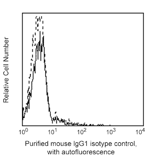
- An isotype control should be used at the same concentration as the antibody of interest.
- Caution: Sodium azide yields highly toxic hydrazoic acid under acidic conditions. Dilute azide compounds in running water before discarding to avoid accumulation of potentially explosive deposits in plumbing.
- For fluorochrome spectra and suitable instrument settings, please refer to our Multicolor Flow Cytometry web page at www.bdbiosciences.com/colors.
- Sodium azide is a reversible inhibitor of oxidative metabolism; therefore, antibody preparations containing this preservative agent must not be used in cell cultures nor injected into animals. Sodium azide may be removed by washing stained cells or plate-bound antibody or dialyzing soluble antibody in sodium azide-free buffer. Since endotoxin may also affect the results of functional studies, we recommend the NA/LE (No Azide/Low Endotoxin) antibody format, if available, for in vitro and in vivo use.
- Please refer to www.bdbiosciences.com/us/s/resources for technical protocols.
Companion Products



.png?imwidth=320)
The 3D3 monoclonal antibody specifically recognizes FcγRII (CD32), a 40 kDa, polymorphic type I transmembrane glycoprotein that serves as a low affinity receptor for aggregated IgG. This highly glycosylated molecule (encoded by at least two different genes) is expressed on monocytes, granulocytes, platelets and B cells. Unlike the FLI28.26 mAb, the 3D3 mAb detected a polymorphic CD32 antigen expressed on B cells of all donors, but only on platelets, monocytes and granulocytes of some donors. The platelets from 3D3+ donors respond to certain stimulatory mAb such as CD165 (clone SN2) which results in aggregation. On the other hand, the platelets from 3D3 negative donors do not form aggregates after stimulation. Individuals can be divided into two groups as responder and non-responder depending on expression, or non-expression, of 3D3. In comparison to the 3D3 mAb, the FLI8.26 mAb detects a monomorphic CD32 antigen expressed on all human donors.
Development References (2)
-
Gosselin EJ, Brown MF, Anderson CL, Zipf TF, Guyre PM. The monoclonal antibody 41H16 detects the Leu 4 responder form of human Fc gamma RII. J Immunol. 1990; 144(5):1817-1822. (Biology). View Reference
-
Vely F, Gruel N, Moncuit J, et al. A new set of monoclonal antibodies against human Fc gamma RII (CD32) and Fc gamma RIII (CD16): characterization and use in various assays.. Hybridoma. 1997; 16(6):519-28. (Immunogen). View Reference
Please refer to Support Documents for Quality Certificates
Global - Refer to manufacturer's instructions for use and related User Manuals and Technical data sheets before using this products as described
Comparisons, where applicable, are made against older BD Technology, manual methods or are general performance claims. Comparisons are not made against non-BD technologies, unless otherwise noted.
For Research Use Only. Not for use in diagnostic or therapeutic procedures.
