





BD Pharmingen™ PerCP-Cy™5.5 Annexin V
(RUO)




Preparation And Storage


Recommended Assay Procedures
PerCP-Cy™5.5 Annexin V is a sensitive probe for identifying apoptotic cells, binding to negatively charged phospholipid surfaces with a higher affinity for phosphatidylserine (PS) than most other phospholipids. PerCP-Cy™5.5 Annexin V binding is calcium dependent and defined calcium and salt concentrations are required for optimal staining as described in the PerCP-Cy™5.5 Annexin V Staining Protocol. Investigators should note that PerCP-Cy™5.5 Annexin V flow cytometric analysis on adherent cell types (eg, HeLa, NIH 3T3, etc.) is not routinely tested as specific membrane damage may occur during cell detachment or harvesting. Methods for utilizing Annexin V for flow cytometry on adherent cell types, however, have been previously reported (Casiola-Rosen et al. and van Engelend et al.).
INDUCTION OF APOPTOSIS BY CAMPTOTHECIN
The following protocol is provided as an illustration on how PerCP-Cy™5.5 Annexin V may be used on a cell line (Jurkat).
Materials
1. Prepare Camptothecin stock solution (Sigma-Aldrich Cat. No. C-9911): 1 mM in DMSO.
2. Jurkat T cells (ATCC TIB-152).
Procedure
1. Add Camptothecin (final conc. 4-6 µM) to 1 × 10^6 Jurkat cells.
2. Incubate the cells for 4-6 hr at 37°C.
3. Proceed with the PerCP-Cy™5.5 Annexin V Staining Protocol to measure apoptosis.
PerCP-Cy™5.5 ANNEXIN V STAINING PROTOCOL
Reagents
1. PerCP-Cy™5.5 Annexin V: Included. Use 5 µl per test.
2. 10X Annexin Binding Buffer: Not Included. 0.1 M Hepes (pH 7.4) 1.4 M NaCl, 25 mM CaCl2. Store at 4°C. Alternatively, BD Pharmingen™ Annexin V Binding Buffer, 10X concentrate (Cat. No. 556454) may be purchased.
Staining
1. Wash cells twice with cold PBS and then resuspend cells in 1× Binding Buffer at a concentration of 1 × 10^6 cells/ml.
2. Transfer 100 µl of the solution (1 × 10^5 cells) to a 5 ml culture tube.
3. Add 5 µl of PerCP-Cy™5.5 Annexin V.
4. Gently vortex the cells and incubate for 15 min at RT (25°C) in the dark.
5. Add 400 µl of 1× Binding Buffer to each tube. Analyze by flow cytometry within 1 hr.
SUGGESTED CONTROLS FOR SETTING UP FLOW CYTOMETRY
The following controls are used to set up markers for quantifying PerCP-Cy™5.5 Annexin V-positive cells:
1. Unstained cells.
2. Cells stained with PerCP-Cy™5.5 Annexin V alone.
Other Staining Controls
A cell line that can be easily induced to undergo apoptosis should be used to obtain positive control staining with PerCP-Cy™5.5 Annexin V. It is important to note that the basal level of apoptosis and necrosis varies considerably within a population. Thus, even in the absence of induced apoptosis, most cell populations will contain a minor percentage of cells that are positive for apoptosis (PerCP-Cy™5.5 Annexin V positive cell).
The untreated population is used to define the basal level of apoptotic and dead cells. The percentage of cells that have been induced to undergo apoptosis is then determined by subtracting the percentage of apoptotic cells in the untreated population from the percentage of apoptotic cells in the treated population.
Product Notices
- Please observe the following precautions: Absorption of visible light can significantly alter the energy transfer occurring in any tandem fluorochrome conjugate; therefore, we recommend that special precautions be taken (such as wrapping vials, tubes, or racks in aluminum foil) to prevent exposure of conjugated reagents, including cells stained with those reagents, to room illumination.
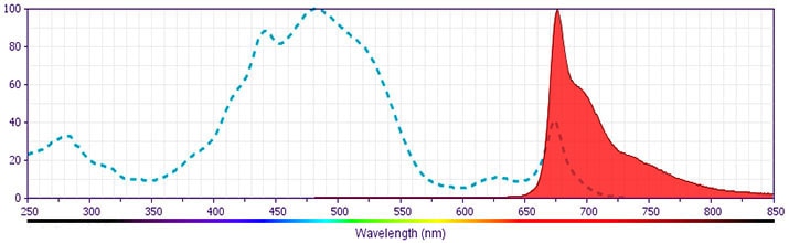
- PerCP-Cy5.5 is optimized for use with a single argon ion laser emitting 488-nm light. Because of the broad absorption spectrum of the tandem fluorochrome, extra care must be taken when using dual-laser cytometers, which may directly excite both PerCP and Cy5.5™. We recommend the use of cross-beam compensation during data acquisition or software compensation during data analysis.
- PerCP-Cy5.5–labelled antibodies can be used with FITC- and R-PE–labelled reagents in single-laser flow cytometers with no significant spectral overlap of PerCP-Cy5.5, FITC, and R-PE fluorescence.
- This product is subject to proprietary rights of Amersham Biosciences Corp. and Carnegie Mellon University and made and sold under license from Amersham Biosciences Corp. This product is licensed for sale only for research. It is not licensed for any other use. If you require a commercial license to use this product and do not have one return this material, unopened to BD Biosciences, 10975 Torreyana Rd, San Diego, CA 92121 and any money paid for the material will be refunded.
- Cy is a trademark of Amersham Biosciences Limited. This conjugated product is sold under license to the following patents: US Patent Nos. 5,486,616; 5,569,587; 5,569,766; 5,627,027.
- Source of all serum proteins is from USDA inspected abattoirs located in the United States.
- Caution: Sodium azide yields highly toxic hydrazoic acid under acidic conditions. Dilute azide compounds in running water before discarding to avoid accumulation of potentially explosive deposits in plumbing.
- For fluorochrome spectra and suitable instrument settings, please refer to our Multicolor Flow Cytometry web page at www.bdbiosciences.com/colors.
- Please refer to www.bdbiosciences.com/us/s/resources for technical protocols.
Companion Products


Apoptosis is a normal physiologic process which occurs during embryonic development as well as in maintenence of tissue homeostasis. The apoptotic program is characterized by certain morphologic features, including loss of plasma membrane asymmetry and attachment, condensation of the cytoplasm and nucleus, and internucleosomal cleavage of DNA. Loss of plasma membrane is one of the earliest features. In apoptotic cells, the membrane phospholipid phosphatidylserine (PS) is translocated from the inner to the outer leaflet of the plasma membrane, thereby exposing PS to the external cellular environment. Annexin V is a 35-36 kDa Ca2+ dependent phospholipid-binding protein that has a high affinity for PS, and binds to cells with exposed PS. Annexin V may be conjugated to fluorochromes including PerCP-Cy™5.5 Annexin V. This format retains its high affinity for PS and thus serves as a sensitive probe for flow cytometric analysis of cells that are undergoing apoptosis. Since externalization of PS occurs in the earlier stages of apoptosis, PerCP-Cy™5.5 Annexin V staining can identify apoptosis at an earlier stage than assays based on nuclear changes such as DNA fragmentation.
NOTE: Investigators should note that the use of Propidium Iodide (PI) or 7-Amino-Actinomycin D (7-AAD) for co-staining with PerCP-Cy5.5 Annexin V is not recommended due to spectral overlap.
Development References (8)
-
Andree HA, Reutelingsperger CP, Hauptmann R, Hemker HC, Hermens WT, Willems GM. Binding of vascular anticoagulant alpha (VAC alpha) to planar phospholipid bilayers. J Biol Chem. 1990; 265(9):4923-4928. (Biology). View Reference
-
Casciola-Rosen L, Rosen A, Petri M, Schlissel M. Surface blebs on apoptotic cells are sites of enhanced procoagulant activity: implications for coagulation events and antigenic spread in systemic lupus erythematosus. Proc Natl Acad Sci U S A. 1996; 93(4):1624-1629. (Methodology: Apoptosis, Flow cytometry). View Reference
-
Homburg CH, de Haas M, von dem Borne AE, Verhoeven AJ, Reutelingsperger CP, Roos D. Human neutrophils lose their surface Fc gamma RIII and acquire Annexin V binding sites during apoptosis in vitro. Blood. 1995; 85(2):532-540. (Biology). View Reference
-
Koopman G, Reutelingsperger CP, Kuijten GA, Keehnen RM, Pals ST, van Oers MH. Annexin V for flow cytometric detection of phosphatidylserine expression on B cells undergoing apoptosis. Blood. 1994; 84(5):1415-1420. (Methodology: Apoptosis, Flow cytometry). View Reference
-
Martin SJ, Reutelingsperger CP, McGahon AJ, et al. Early redistribution of plasma membrane phosphatidylserine is a general feature of apoptosis regardless of the initiating stimulus: inhibition by overexpression of Bcl-2 and Abl. J Exp Med. 1995; 182(5):1545-1556. (Biology). View Reference
-
Raynal P, Pollard HB. Annexins: the problem of assessing the biological role for a gene family of multifunctional calcium- and phospholipid-binding proteins. Biochim Biophys Acta. 1994; 1197(1):63-93. (Biology). View Reference
-
Vermes I, Haanen C, Steffens-Nakken H, Reutelingsperger C. A novel assay for apoptosis. Flow cytometric detection of phosphatidylserine expression on early apoptotic cells using fluorescein labelled Annexin V. J Immunol Methods. 1995; 184(1):39-51. (Methodology: Apoptosis, Flow cytometry). View Reference
-
van Engeland M, Ramaekers FC, Schutte B, Reutelingsperger CP. A novel assay to measure loss of plasma membrane asymmetry during apoptosis of adherent cells in culture. Cytometry. 1996; 24(2):131-139. (Methodology: Apoptosis, Flow cytometry). View Reference
Please refer to Support Documents for Quality Certificates
Global - Refer to manufacturer's instructions for use and related User Manuals and Technical data sheets before using this products as described
Comparisons, where applicable, are made against older BD Technology, manual methods or are general performance claims. Comparisons are not made against non-BD technologies, unless otherwise noted.
For Research Use Only. Not for use in diagnostic or therapeutic procedures.
