







Preparation And Storage


Recommended Assay Procedures
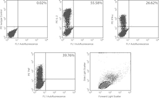
Immunofluorescent Staining and Flow Cytometric Analysis: The PE-conjugated B27 antibody is useful for multicolor immunofluorescent staining and flow cytometric analysis to identify and enumerate IFN-γ producing cells within mixed cell populations (see image). This 100 Test Size formulation of the PE-conjugated B27 antibody has been pre-titrated to assure effective intracellular detection of human IFN-γ using 20 µl per 1 x 10e6 cells. For specific methodology, please visit our website, http://www.bdbiosciences.com/us/s/resources and refer to the protocols section under "Cytokines (Intracellular Staining)" or "Intracellular Flow".
A suitable mouse IgG1 isotype control for assessing the level of background staining on paraformaldehyde-fixed/saponinpermeabilized
human cells is also available in a 100 Test Size formulation PE-MOPC-21 (Cat. No. 559320). A useful control for demonstrating specificity of staining is the following: pre-block the paraformaldehyde-fixed/saponin-permeabilized cells with unlabeled B27 antibody (Cat. No. 554699/550011) prior to staining. The intracellular cytokine staining technique and blocking controls are described in detail by C. Prussin and D. Metcalfe.
Important Note: This pre-titered antibody solution does not contain a cell permeabilization agent. It is necessary to include a cell permeabilization agent when using the pre-titered antibody solution to stain fixed and permeabilized cells. Perm/Wash™ Buffer (Cat. No. 554723) contains the permeabilization agent saponin and is useful for this purpose as described in the USAGE section below.
USAGE
1. Resuspend 1 x 10^6 fixed and permeabilized cells in 20 µl of the pre-titered antibody solution and 30 µl of 1X Perm/Wash™ Buffer.
2. Incubate the cell suspension for 15 minutes (4°C, in the dark).
3. Wash twice in 100 µl of 1X Perm/Wash™ Buffer.
Product Notices
- This reagent has been pre-diluted for use at the recommended Volume per Test. We typically use 1 × 10^6 cells in a 100-µl experimental sample (a test).
- An isotype control should be used at the same concentration as the antibody of interest.
- Source of all serum proteins is from USDA inspected abattoirs located in the United States.
- Caution: Sodium azide yields highly toxic hydrazoic acid under acidic conditions. Dilute azide compounds in running water before discarding to avoid accumulation of potentially explosive deposits in plumbing.
- For fluorochrome spectra and suitable instrument settings, please refer to our Multicolor Flow Cytometry web page at www.bdbiosciences.com/colors.
- Species cross-reactivity detected in product development may not have been confirmed on every format and/or application.
- Please refer to www.bdbiosciences.com/us/s/resources for technical protocols.
Companion Products




The B27 monoclonal antibody specifically binds to human interferon-γ (IFN-γ), a 14-18 kDa glycoprotein containing 143 amino acid residues. IFN-γ is a potent multifunctional cytokine produced by several activated cell types including NK, NKT, CD4+TCRαβ+, CD8+TCRαβ+, and TCRγδ+ T cells. IFN-γ exerts its biological effects through specific binding to the high-affinity IFN-γ receptor complex comprised of IFN-γRα (CD119) and IFN-γRβ subunits. In addition to its antiviral effects, IFN-γ upregulates a number of lymphoid cell functions including the antimicrobial and anti-tumor responses of macrophages, NK cells, and neutrophils. In addition, IFN-γ influences the regulation of proliferation, differentiation, and effector responses of B cell and T cell subsets. These influences can involve IFN-γ's capacity to boost MHC class I and II expression by antigen-presenting cells as well as direct effects on B cells and T cells themselves. B27 is a neutralizing antibody. The use of B27 antibody for epitope mapping of human IFN-γ has been described. The B27 antibody has been reported not to bind to denatured IFN-γ.

Development References (5)
-
Abrams JS, Roncarolo MG, Yssel H, Andersson U, Gleich GJ, Silver JE. Strategies of anti-cytokine monoclonal antibody development: immunoassay of IL-10 and IL-5 in clinical samples. Immunol Rev. 1992; 127:5-24. (Clone-specific). View Reference
-
Favre C, Wijdenes J, Cabrillat H, Djossou O, Banchereau J, de Vries JE. Epitope mapping of recombinant human gamma interferon using monoclonal antibodies. Mol Immunol. 1989; 26(1):17-25. (Clone-specific: Immunoprecipitation, Neutralization). View Reference
-
Fonteneau JF, Le Drean E, Le Guiner S, Gervois N, Diez E, Jotereau F. Heterogeneity of biologic responses of melanoma-specific CTL. J Immunol. 1997; 159(6):2831-2839. (Biology). View Reference
-
Prussin C, Metcalfe DD. Detection of intracytoplasmic cytokine using flow cytometry and directly conjugated anti-cytokine antibodies. J Immunol Methods. 1995; 188(1):117-128. (Methodology: Flow cytometry). View Reference
-
Rotteveel FT, Kokkelink I, van Lier RA, et al. Clonal analysis of functionally distinct human CD4+ T cell subsets. J Exp Med. 1988; 168(5):1659-1673. (Biology). View Reference
Please refer to Support Documents for Quality Certificates
Global - Refer to manufacturer's instructions for use and related User Manuals and Technical data sheets before using this products as described
Comparisons, where applicable, are made against older BD Technology, manual methods or are general performance claims. Comparisons are not made against non-BD technologies, unless otherwise noted.
For Research Use Only. Not for use in diagnostic or therapeutic procedures.
