







Preparation And Storage


Product Notices
- Since applications vary, each investigator should titrate the reagent to obtain optimal results.
- An isotype control should be used at the same concentration as the antibody of interest.
- Caution: Sodium azide yields highly toxic hydrazoic acid under acidic conditions. Dilute azide compounds in running water before discarding to avoid accumulation of potentially explosive deposits in plumbing.
- Source of all serum proteins is from USDA inspected abattoirs located in the United States.
- Pacific Blue™ is a trademark of Molecular Probes, Inc., Eugene, OR.
- Brilliant Violet™ 421 is a trademark of Sirigen.
- For fluorochrome spectra and suitable instrument settings, please refer to our Multicolor Flow Cytometry web page at www.bdbiosciences.com/colors.
- Please refer to www.bdbiosciences.com/us/s/resources for technical protocols.
Companion Products




.png?imwidth=320)
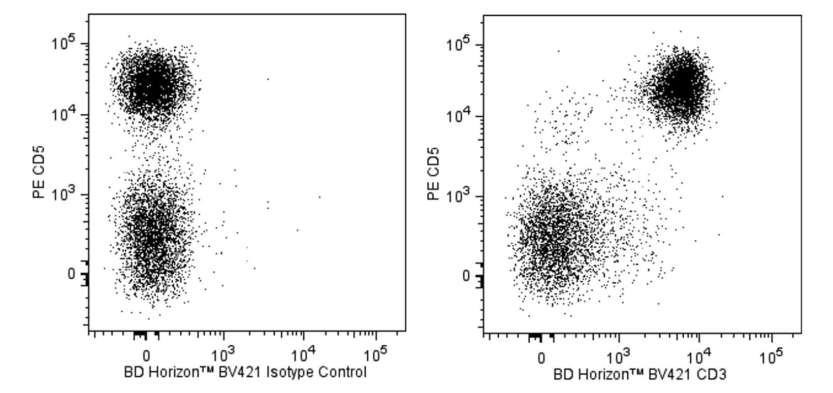
The 1F4 antibody reacts with the T-cell receptor-associated CD3 cell-surface antigen found on thymocytes, peripheral T lymphocytes, and dendritic epidermal T cells. In vitro, immobilized 1F4 mAb induces T-cell proliferation, and soluble mAb synergizes with PMA for T-cell activation. Pre-incubation of splenocytes with mAb 1F4 blocks staining with mAb G4.18 (Cat. No. 559974/554830).
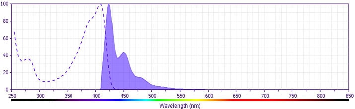
The antibody was conjugated to BD Horizon™ BV421 which is part of the BD Horizon Brilliant Violet™ family of dyes. With an Ex Max of 407-nm and Em Max at 421-nm, BD Horizon BV421 can be excited by the violet laser and detected in the standard Pacific Blue™ filter set (eg, 450/50-nm filter). BD Horizon BV421 conjugates are very bright, often exhibiting a 10 fold improvement in brightness compared to Pacific Blue™ conjugates.
Development References (3)
-
Elbe A, Kilgus O, Hünig T, and Stingl G. T-cell receptor diversity in dendritic epidermal T cells in the rat. J Invest Dermatol. 1993; 102:74-79. (Biology). View Reference
-
Nicolls MR, Aversa GG, Pearce NW, et al. Induction of long-term specific tolerance to allografts in rats by therapy with an anti-CD3-like monoclonal antibody.. Transplantation. 1993; 55(3):459-68. (Biology). View Reference
-
Tanaka T, Masuko T, Yagita H, Tamura T, Hashimoto Y. Characterization of a CD3-like rat T cell surface antigen recognized by a monoclonal antibody.. J Immunol. 1989; 142(8):2791-2795. (Immunogen: Flow cytometry, Functional assay, Immunohistochemistry, Stimulation). View Reference
Please refer to Support Documents for Quality Certificates
Global - Refer to manufacturer's instructions for use and related User Manuals and Technical data sheets before using this products as described
Comparisons, where applicable, are made against older BD Technology, manual methods or are general performance claims. Comparisons are not made against non-BD technologies, unless otherwise noted.
For Research Use Only. Not for use in diagnostic or therapeutic procedures.
