Skip to main content Skip to navigation
Alexa Fluor® 647 Mouse anti-Human CCL18/DC-CK1
Flow cytometric analysis of human CCL18 expression in cultured immature monocyte-derived dendritic cells or unstimulated peripheral blood monocytes. Immature monocyte-derived dendritic cells (MoDC; Left Panel) were generated from peripheral blood monocytes cultured in complete Iscove's Modified Dulbecco's Medium (IMDM) containing recombinant human IL-4 (Cat. No. 554605) and GM-CSF (Cat. No. 550068) for 5 days. Peripheral blood monocytes (PB-M; Right Panel) from freshly-isolated peripheral blood mononuclear cells served as control cells. Cells were fixed and permeabilized using BD Cytofix/Cytoperm™ Fixation/Permablization Kit reagents (Cat. No. 554714) followed by staining with Alexa Fluor® 647 Mouse Anti-Human CCL18 antibody (Cat. No. 561318). Histograms show the expression pattern of CCL18 (solid line histogram) or an Alexa Fluor® 647 Mouse IgG1, κ Isotype Control (Cat No. 557732, dashed line histogram) in these two cell populations derived by gating events with the forward and side light-scatter characteristics of intact MoDC or PB-M. Flow cytometry was performed using a BD™ LSR II Flow Cytometry System.
Product Details
Down Arrow Up Arrow


BD Pharmingen™
C-C motif chemokine 18; Dendritic cell chemokine 1; MIP-4; PARC; AMAC-1
Human (QC Testing)
Mouse IgG1, κ
Human CCL18 Recombinant Protein
Intracellular staining (flow cytometry) (Routinely Tested)
5 µl
6362
AB_10898008
Aqueous buffered solution containing BSA, protein stabilizer, and ≤0.09% sodium azide.
RUO


Preparation And Storage

Store undiluted at 4°C and protected from prolonged exposure to light. Do not freeze. The monoclonal antibody was purified from tissue culture supernatant or ascites by affinity chromatography. The antibody was conjugated to Alexa Fluor® 647 under optimum conditions, and unreacted Alexa Fluor® 647 was removed.

Product Notices

  1. Source of all serum proteins is from USDA inspected abattoirs located in the United States.
  2. This reagent has been pre-diluted for use at the recommended Volume per Test. We typically use 1 × 10^6 cells in a 100-µl experimental sample (a test).
  3. The Alexa Fluor®, Pacific Blue™, and Cascade Blue® dye antibody conjugates in this product are sold under license from Molecular Probes, Inc. for research use only, excluding use in combination with microarrays, or as analyte specific reagents. The Alexa Fluor® dyes (except for Alexa Fluor® 430), Pacific Blue™ dye, and Cascade Blue® dye are covered by pending and issued patents.
  4. Alexa Fluor® is a registered trademark of Molecular Probes, Inc., Eugene, OR.
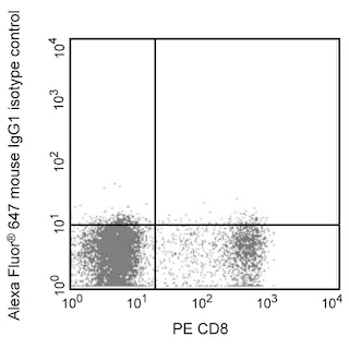
  5. Alexa Fluor® 647 fluorochrome emission is collected at the same instrument settings as for allophycocyanin (APC).
  6. Caution: Sodium azide yields highly toxic hydrazoic acid under acidic conditions. Dilute azide compounds in running water before discarding to avoid accumulation of potentially explosive deposits in plumbing.
  7. For fluorochrome spectra and suitable instrument settings, please refer to our Multicolor Flow Cytometry web page at www.bdbiosciences.com/colors.
  8. An isotype control should be used at the same concentration as the antibody of interest.
  9. Please refer to www.bdbiosciences.com/us/s/resources for technical protocols.
561318 Rev. 1
Antibody Details
Down Arrow Up Arrow
AZN-CK18

The AZN-CK18 monoclonal antibody specifically binds to the CC chemokine ligand 18 (CCL18), also known as Dendritic cell chemokine 1 (DC-CK1). CCL18 is expressed by dendritic cells and activated (eg, with LPS or cytokines including IL-4, IL-10, IL-13) monocytes and macrophages. CCL18 is a chemotactic factor that primarily attracts T and B lymphocytes (but not monocytes or granulocytes) towards dendritic cells and macrophages. This suggests that CCL18 likely plays roles in the development of both humoral and cell-mediated immune responses and may participate is certain inflammatory diseases, such as those affecting the joints and lungs.


561318 Rev. 1
Format Details
Down Arrow Up Arrow
Alexa Fluor™ 647
Alexa Fluor™ 647 Dye is part of the BD red family of dyes. This is a small organic fluorochrome with an excitation maximum (Ex Max) at 653-nm and an emission maximum (Em Max) at 669-nm. Alexa Fluor 647 is designed to be excited by the Red laser (627-640 nm) and detected using an optical filter centered near 520-nm (e.g., a 660/20 nm bandpass filter). Please ensure that your instrument’s configurations (lasers and optical filters) are appropriate for this dye.
Alexa Fluor™ 647
Red 627-640 nm
653 nm
669 nm
561318 Rev.1
Citations & References
Down Arrow Up Arrow
View product citations for antibody "561318" on CiteAb

Development References (3)

  1. Lindhout E, Vissers JL, Hartgers FC, et al. The dendritic cell-specific CC-chemokine DC-CK1 is expressed by germinal center dendritic cells and attracts CD38-negative mantle zone B lymphocytes. J Immunol. 2001; 166(5):3284-3289. (Clone-specific: Immunohistochemistry). View Reference
  2. Schutyser E., Richmond A., and Van Damme J. Involvement of CC chemokine ligand 18(CCL18) in normal and pathological process. J Leukoc Biol. 2005; 78(1):14-26. (Biology). View Reference
  3. van der Voort R, Kramer M, Lindhout E, et al. Novel monoclonal antibodies detect elevated levels of the chemokine CCL18/DC-CK1 in serum and body fluids in pathological conditions. J Leukoc Biol. 2005; 77(5):739-747. (Immunogen). View Reference
561318 Rev. 1

Please refer to Support Documents for Quality Certificates


Global - Refer to manufacturer's instructions for use and related User Manuals and Technical data sheets before using this products as described


Comparisons, where applicable, are made against older BD Technology, manual methods or are general performance claims.  Comparisons are not made against non-BD technologies, unless otherwise noted.

For Research Use Only. Not for use in diagnostic or therapeutic procedures.