Old Browser
This page has been recently translated and is available in French now.
Looks like you're visiting us from {countryName}.
Would you like to stay on the current country site or be switched to your country?


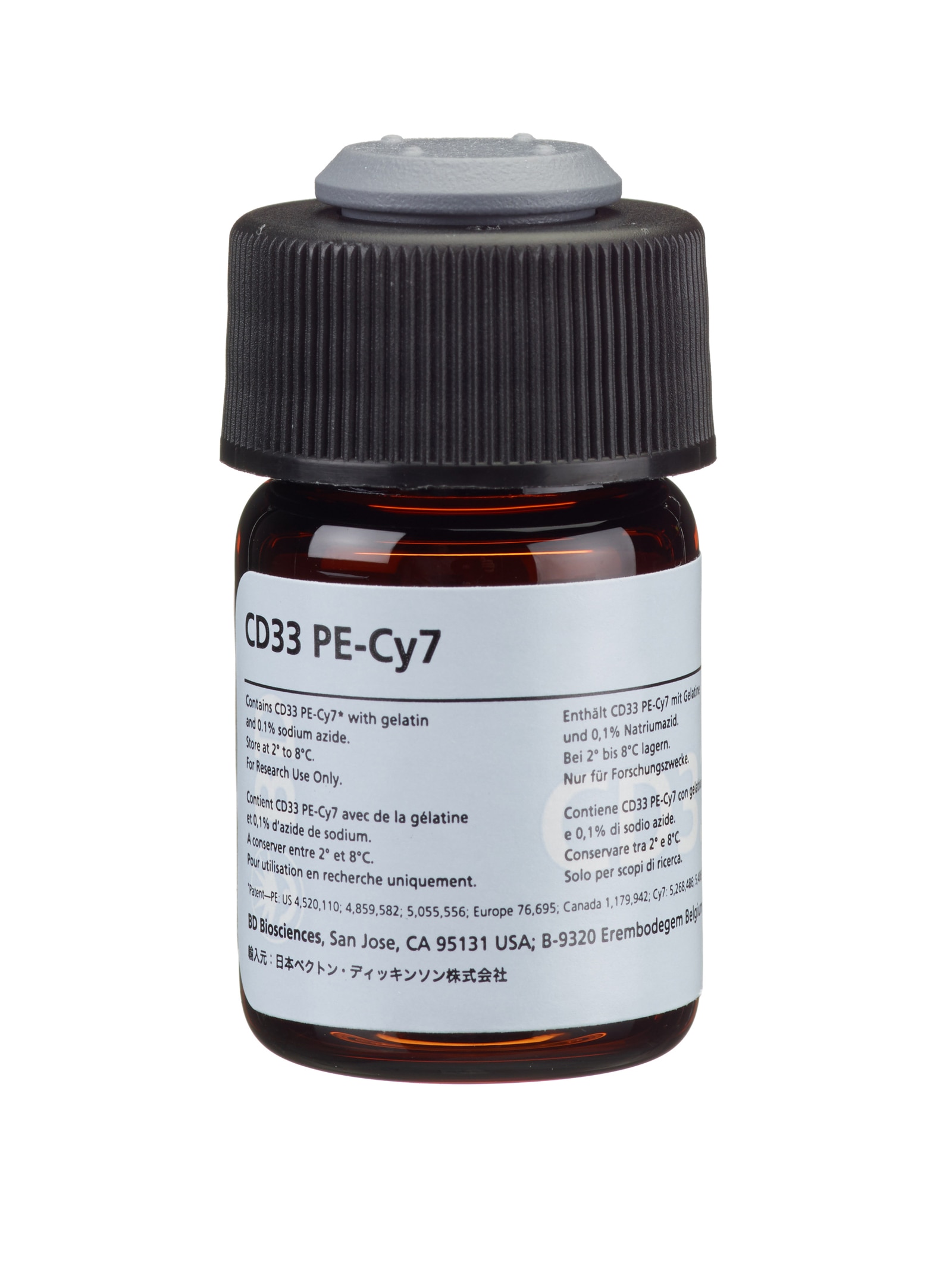
PE-Cy™7 Mouse Anti-Human CD33
Regulatory Status Legend
Any use of products other than the permitted use without the express written authorization of Becton, Dickinson and Company is strictly prohibited.
Preparation And Storage
The monoclonal antibody is supplied as 12 μg purified immunoglobulin in 2.0 mL (6 μg/mL) of phosphate-buffered saline (PBS). The FITC conjugate is supplied as 3 μg in 1.0 mL (3 μg/mL) of PBS. The PE conjugate is supplied as 24 μg in 2.0 mL (12 μg/mL) of PBS. The PerCP-Cy5.5 conjugate is supplied as 6.3 μg in 1.0 mL (6.3 μg/mL) of PBS. The PE-Cy7 conjugate is supplied as 12.5 μg in 0.5 mL (25 μg/mL) of PBS. The APC conjugate is supplied as 6.2 μg in 0.5 mL (12.4 μg/mL) of PBS. PBS contains gelatin and 0.1% sodium azide.
Store vials at 2° to 8°C. Conjugated forms should not be frozen and should be protected from prolonged exposure to light. Each reagent is stable for the period shown on the bottle label when stored as directed.
The CD33 antibody, clone P67-6, is derived from the hybridization of Sp2/0 mouse myeloma cells with spleen cells isolated from BALB/c mice immunized with FMY9S5 cells containing the CD33 gene. The CD33 antibody recognizes a human myelomonocytic antigen, with a molecular weight of 67 kilodaltons (kDa).

Development References (7)
-
Andrews RG, Takahashi M, Segal GM, Powell JS, Bernstein ID, Singer JW. The L4F3 antigen is expressed by unipotent and multipotent colony-forming cells but not by their precursors. Blood. 1986; 68:1030-1035. (Biology).
-
Andrews RG, Torok-Storb B, Bernstein ID. Myeloid-associated differentiation antigens on stem cells and their progeny identified by monoclonal antibodies.. Blood. 1983; 62(1):124-32. (Biology). View Reference
-
Bernstein ID, Singer JW, Andrews RG, et al. Treatment of acute myeloid leukemia cells in vitro with a monoclonal antibody recognizing a myeloid differentiation antigen allows normal progenitor cells to be expressed.. J Clin Invest. 1987; 79(4):1153-9. (Biology). View Reference
-
Dinndorf PA, Andrews RG, Benjamin D, Ridgway D, Wolff L, Bernstein ID. Expression of normal myeloid-associated antigens by acute leukemia cells.. Blood. 1986; 67(4):1048-53. (Biology). View Reference
-
Foon KA, Todd RF. Immunologic classification of leukemia and lymphoma.. Blood. 1986; 68(1):1-31. (Biology). View Reference
-
Köller U, Peschel CH. Knapp W, Dörken B, Gilks WR, et al, ed. Leucocyte Typing IV: White Cell Differentiation Antigens. New York, NY: Oxford University Press; 1989:812-813.
-
Terstappen LW, Hollander Z, Meiners H, Loken MR. Quantitative comparison of myeloid antigens on five lineages of mature peripheral blood cells. J Leukoc Biol. 1990; 48(2):138-148. (Biology). View Reference
Please refer to Support Documents for Quality Certificates
Global - Refer to manufacturer's instructions for use and related User Manuals and Technical data sheets before using this products as described
Comparisons, where applicable, are made against older BD Technology, manual methods or are general performance claims. Comparisons are not made against non-BD technologies, unless otherwise noted.
For Research Use Only. Not for use in diagnostic or therapeutic procedures.
Although not required, these products are manufactured in accordance with Good Manufacturing Practices.