Dear Customer, If you experience any text readability or display issues on our website, please update your web browser to the latest version
.
See More
Dear Customer, If you experience any text readability or display issues on our website, please update your web browser to the latest version
.
See MoreDear Customer, If you experience any text readability or display issues on our website, please update your web browser to the latest version
.
Looks like you're visiting us from {countryName}.
Would you like to stay on the current location site or be switched to your location?
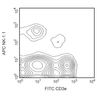
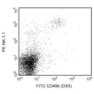
Flow cytometric analysis of PE anti-mouse CD335 (NKp46) expression on mouse splenocytes. C57BL/6 and BALB/c mouse spleen cells were stained separately with PE anti-mouse CD335 (NKp46) antibody. After washing, C57BL/6 cells were stained with APC-conjugated anti-mouse NK-1.1 (NKR-P1B and NKR-P1C) antibody (Cat. No.550627; left panel) and BALB/c cells were stained with FITC-conjugated anti-mouse CD49b (DX5) antibody (Cat. No.553857; right panel). Two-color dot plots showing the correlated expression patterns of CD335/NKp46 and either NK-1.1/CD161 (C57BL/6 cells; left panel) or DX5/CD49b (BALB/c cells; right panel) were derived from gated events with the forward and side light-scatter characteristics of viable lymphocytes. Flow cytometry was performed using a BD™ LSRII System.
Any use of products other than the permitted use without the express written authorization of Becton, Dickinson and Company is strictly prohibited.
The monoclonal antibody 29A1.4 specifically binds to mouse CD335, also known as NKp46. NKp46 is a 46 kDa type I transmembrane glycoprotein that is a member of the natural cytotoxicity receptor (NCR) family and immunoglobulin superfamily. NKp46 is encoded by the Ncr1 gene located on chromosome 7. NKp46 functions as a cytotoxicity triggering receptor and is selectively expressed by immature and mature NK cells in all mouse strains tested. NKp46 is detected on a minute fraction of NK-like T cells (less than 2% of NKp46+ express CD3e) but not on CD1d-restricted NKT cells from C57BL/6 mice. When immobilized on tissue culture plates, the 29A1.4 antibody reportedly stimulates NK cells to produce interferon-gamma and to release their cytoplasmic granule contents. Although the ligands for the NKp46 receptor have not been fully characterized, recent evidence indicates that this receptor plays an important role in the NK cell-mediated recognition and killing of some virus-infected cells and tumor cells. The immunogen used to generate the 29A1.4 clone was mouse NKp46-Fc recombinant protein.

Please refer to Support Documents for Quality Certificates
Global - Refer to manufacturer's instructions for use and related User Manuals and Technical data sheets before using this products as described
Comparisons, where applicable, are made against older BD Technology, manual methods or are general performance claims. Comparisons are not made against non-BD technologies, unless otherwise noted.
For Research Use Only. Not for use in diagnostic or therapeutic procedures.
Refer to manufacturer's instructions for use and related User Manuals and Technical Data Sheets before using this product as described.
Comparisons, where applicable, are made against older BD technology, manual methods or are general performance claims. Comparisons are not made against non-BD technologies, unless otherwise noted.