





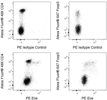
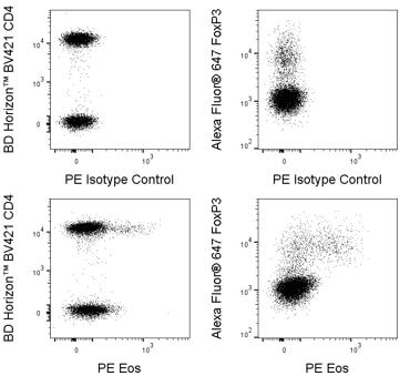
.png)


Preparation And Storage


Product Notices
- Since applications vary, each investigator should titrate the reagent to obtain optimal results.
- An isotype control should be used at the same concentration as the antibody of interest.
- Caution: Sodium azide yields highly toxic hydrazoic acid under acidic conditions. Dilute azide compounds in running water before discarding to avoid accumulation of potentially explosive deposits in plumbing.
- For fluorochrome spectra and suitable instrument settings, please refer to our Multicolor Flow Cytometry web page at www.bdbiosciences.com/colors.
- Alexa Fluor® is a registered trademark of Molecular Probes, Inc., Eugene, OR.
- Species cross-reactivity detected in product development may not have been confirmed on every format and/or application.
- Please refer to www.bdbiosciences.com/us/s/resources for technical protocols.
Companion Products


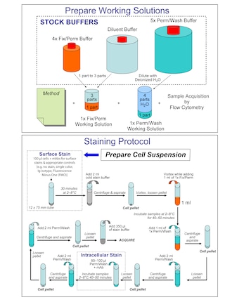
.png?imwidth=320)
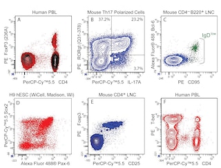
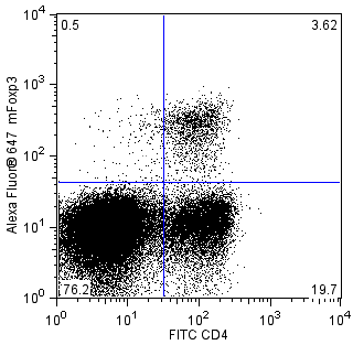
The W7-486 monoclonal antibody specifically recognizes mouse and human Eos. Eos is a member of the Ikaros family of zinc-finger transcription factors that play important roles in hematopoietic cell development and tumor suppression. Other members of the Ikaros family include Ikaros, Helios, Aiolos, and Pegasus. Eos is encoded by the IKAROS family zinc finger 4 (mouse Ikzf4 and human IKZF4) genes. This DNA-binding transcription factor forms homodimers and heterodimers with other Ikaros family members. In the peripheral immune system, Eos expression is predominantly in CD4-positive FoxP3-positive Treg cells, where it is involved in the negative regulation of gene expression. It has been reported that Eos is co-expressed with Helios, but not with Aiolos, in Treg cells. Upon in vitro activation, FoxP3-negative, CD4- or CD8-positive T lymphocytes may express Eos.

Development References (3)
-
Pan F, Yu H, Dang EV, et al. Eos mediates Foxp3-dependent gene silencing in CD4+ regulatory T cells. Science. 2009; 325(5944):1142-1146. (Biology). View Reference
-
Raffin C, Pignon P, Celse C, Debien E, Valmori D, Ayyoub M. Human memory Helios- FOXP3+ regulatory T cells (Tregs) encompass induced Tregs that express Aiolos and respond to IL-1β by downregulating their suppressor functions. J Immunol. 2013; 191(9):4619-27. (Biology). View Reference
-
Rieder SA, Metidji A, Glass DD, et al. Eos Is Redundant for Regulatory T Cell Function but Plays an Important Role in IL-2 and Th17 Production by CD4+ Conventional T Cells. J Immunol. 2015; 195(2):553-63. (Biology). View Reference
Please refer to Support Documents for Quality Certificates
Global - Refer to manufacturer's instructions for use and related User Manuals and Technical data sheets before using this products as described
Comparisons, where applicable, are made against older BD Technology, manual methods or are general performance claims. Comparisons are not made against non-BD technologies, unless otherwise noted.
For Research Use Only. Not for use in diagnostic or therapeutic procedures.
Refer to manufacturer's instructions for use and related User Manuals and Technical Data Sheets before using this product as described.
Comparisons, where applicable, are made against older BD technology, manual methods or are general performance claims. Comparisons are not made against non-BD technologies, unless otherwise noted.
