





.png)


Preparation And Storage


Recommended Assay Procedures
The PE-conjugated 11A9 antibody (Cat. No. 559562/561019/551773) can be used for the immunofluorescent staining and flow cytometric analyses of human leukocytes (see image) and cell lines that express CCR6. Please titrate between 0.1 and 1.0 µg/test.
Product Notices
- Since applications vary, each investigator should titrate the reagent to obtain optimal results.
- An isotype control should be used at the same concentration as the antibody of interest.
- Caution: Sodium azide yields highly toxic hydrazoic acid under acidic conditions. Dilute azide compounds in running water before discarding to avoid accumulation of potentially explosive deposits in plumbing.
- For fluorochrome spectra and suitable instrument settings, please refer to our Multicolor Flow Cytometry web page at www.bdbiosciences.com/colors.
- Species cross-reactivity detected in product development may not have been confirmed on every format and/or application.
- Please refer to www.bdbiosciences.com/us/s/resources for technical protocols.
Companion Products


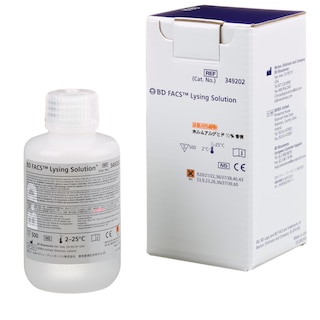

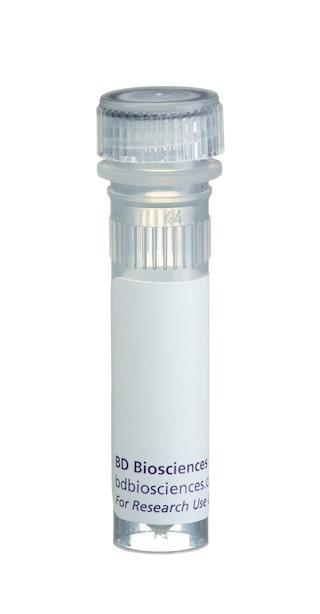
The 11A9 monoclonal antibody specifically binds to CD196, which is also known as CCR6. CCR6 is a seven-transmembrane, G-protein-coupled, glycoprotein receptor that is a member of the beta chemokine receptor family. The human CCR6 gene has been mapped to chromosome 6q27. CCR6 is a receptor for the CC chemokine CCL20/MIP-3alpha/LARC/Exodus and also binds with lower affinity to and mediates responses to beta-defensin2/hBD-2. CCR6 is predominantly expressed by B lymphocytes, certain subsets of effector and memory T cells and by immature dendritic cells but not by monocytes, NK cells, or granulocytes. Skin-homing CLA (Cutaneous Lymphocyte Antigen)-positive memory T cells, Th1 cells, regulatory T cells and IL-17A-producing Th17 cells predominantly express high levels of CCR6. CCR6 mediates the trafficking of T, B, and dendritic cells to epithelial sites near the skin and mucosal surfaces during inflammatory and immunological responses. An N-terminal peptide of human CCR6 was used as an immunogen to generate the 11A9 hybridoma. The 11A9 antibody does not cross-react with human CCR1, CCR2, CCR3, CCR4, CCR5, CCR7, CCR8, CCR9, CXCR1, CXCR2, CXCR3, CXCR4 and CXCR5 receptors. This antibody is NOT a neutralizing antibody.

Development References (7)
-
Baba M, Imai T, Nishimura M, et al. Identification of CCR6, the specific receptor for a novel lymphocyte-directed CC chemokine LARC. J Biol Chem. 1997; 272(23):14893-14898. (Biology). View Reference
-
Greaves DR, Wang W, Dairaghi DJ, et al. CCR6, a CC chemokine receptor that interacts with macrophage inflammatory protein 3alpha and is highly expressed in human dendritic cells. J Exp Med. 1997; 186(6):837-844. (Biology). View Reference
-
Liao F, Alderson R, Su J, Ullrich SJ, Kreider BL, Farber JM. STRL22 is a receptor for the CC chemokine MIP-3alpha. Biochem Biophys Res Commun. 1997; 236(1):212-217. (Biology). View Reference
-
Liao F, Lee HH, Farber JM. Cloning of STRL22, a new human gene encoding a G-protein-coupled receptor related to chemokine receptors and located on chromosome 6q27. Genomics. 1997; 40(1):175-180. (Biology). View Reference
-
Liao F, Rabin RL, Smith CS, Sharma G, Nutman TB, Farber JM. CC-chemokine receptor 6 is expressed on diverse memory subsets of T cells and determines responsiveness to macrophage inflammatory protein 3 alpha. J Immunol. 1999; 162(1):186-194. (Biology). View Reference
-
Power CA, Church DJ, Meyer A, et al. Cloning and characterization of a specific receptor for the novel CC chemokine MIP-3alpha from lung dendritic cells. J Exp Med. 1997; 186(6):825-835. (Biology). View Reference
-
Zaballos A, Varona R, Gutierrez J, Lind P, Marquez G. Molecular cloning and RNA expression of two new human chemokine receptor-like genes. Biochem Biophys Res Commun. 1996; 227(3):846-853. (Biology). View Reference
Please refer to Support Documents for Quality Certificates
Global - Refer to manufacturer's instructions for use and related User Manuals and Technical data sheets before using this products as described
Comparisons, where applicable, are made against older BD Technology, manual methods or are general performance claims. Comparisons are not made against non-BD technologies, unless otherwise noted.
For Research Use Only. Not for use in diagnostic or therapeutic procedures.
Refer to manufacturer's instructions for use and related User Manuals and Technical Data Sheets before using this product as described.
Comparisons, where applicable, are made against older BD technology, manual methods or are general performance claims. Comparisons are not made against non-BD technologies, unless otherwise noted.
