







Preparation And Storage


Recommended Assay Procedures
BD™ CompBeads can be used as surrogates to assess fluorescence spillover (Compensation). When fluorochrome conjugated antibodies are bound to CompBeads, they have spectral properties very similar to cells. However, for some fluorochromes there can be small differences in spectral emissions compared to cells, resulting in spillover values that differ when compared to biological controls. It is strongly recommended that when using a reagent for the first time, users compare the spillover on cells and CompBead to ensure that BD Comp beads are appropriate for your specific cellular application.
For optimal and reproducible results, BD Horizon Brilliant Stain Buffer should be used anytime two or more BD Horizon Brilliant dyes are used in the same experiment. Fluorescent dye interactions may cause staining artifacts which may affect data interpretation. The BD Horizon Brilliant Stain Buffer was designed to minimize these interactions. More information can be found in the Technical Data Sheet of the BD Horizon Brilliant Stain Buffer (Cat. No. 563794/566349) or the BD Horizon Brilliant Stain Buffer Plus (Cat. No. 566385).
For optimal results, it is recommended to perform 2 washes after staining with antibodies. Cells may be prepared, stained with antibodies and washed twice with wash buffer per established protocols for immunofluorescence staining, prior to acquisition on a flow cytometer. Performing fewer than the recommended wash steps may lead to increased spread of the negative population.
Product Notices
- This reagent has been pre-diluted for use at the recommended Volume per Test. We typically use 1 × 10^6 cells in a 100-µl experimental sample (a test).
- An isotype control should be used at the same concentration as the antibody of interest.
- Caution: Sodium azide yields highly toxic hydrazoic acid under acidic conditions. Dilute azide compounds in running water before discarding to avoid accumulation of potentially explosive deposits in plumbing.
- For fluorochrome spectra and suitable instrument settings, please refer to our Multicolor Flow Cytometry web page at www.bdbiosciences.com/colors.
- BD Horizon Brilliant Stain Buffer is covered by one or more of the following US patents: 8,110,673; 8,158,444; 8,575,303; 8,354,239.
- Please refer to http://regdocs.bd.com to access safety data sheets (SDS).
- Please refer to www.bdbiosciences.com/us/s/resources for technical protocols.
Companion Products




The SK7 (Leu-4) monoclonal antibody specifically binds to the epsilon chain of the CD3 antigen/T-cell antigen receptor (TCR) complex. This complex is composed of at least six proteins that range in molecular weight from 20 to 30 kDa. The antigen recognized by CD3 antibodies is noncovalently associated with either α/β or γ/δ TCR (70 to 90 kDa). The CD3 antigen is present on 61% to 85% of normal peripheral blood lymphocytes 60% to 85% of thymocytes and on Purkinje cells in the cerebellum. The soluble form of this antibody has a mitogenic effect on most peripheral blood T lymphocytes, provided appropriate functional monocytes are present.
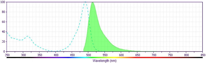
The antibody was conjugated to BD Horizon BB515 which is part of the BD Horizon Brilliant™ Blue family of dyes. With an Ex Max near 490 nm and an Em Max near 515 nm, BD Horizon BB515 can be excited by the blue laser (488 nm) laser and detected with a 530/30 nm filter. This dye has been exclusively developed by BD Biosciences and is up to seven times brighter than FITC with less spillover into the PE channel. Due to similar excitation and emission properties, BB515, FITC, and Alexa Fluor® 488 cannot be used simultaneously. It is not recommended to use BB515 in cocktails that include Streptavidin conjugates as it may cause high background.
Development References (13)
-
Ernst DN, Shih CC. CD3 complex. J Biol Regul Homeost Agents. 2000; 14(3):226-229. (Biology). View Reference
-
Kan EA, Wang CY, Wang LC, Evans RL. Noncovalently bonded subunits of 22 and 28 kd are rapidly internalized by T cells reacted with anti-Leu-4 antibody. J Immunol. 1983; 131(2):536-539. (Clone-specific: Flow cytometry, Functional assay, Immunofluorescence, Immunoprecipitation). View Reference
-
Kaneoka H, Perez-Rojas G, Sasasuki T, Benike CJ, Engleman EG. Human T lymphocyte proliferation induced by a pan-T monoclonal antibody (anti-Leu 4): heterogeneity of response is a function of monocytes. J Immunol. 1983; 131(1):158-164. (Clone-specific: Activation, Functional assay, Stimulation). View Reference
-
Knapp W. W. Knapp .. et al., ed. Leucocyte typing IV : white cell differentiation antigens. Oxford New York: Oxford University Press; 1989:1-1182.
-
Knowles RW. Immunochemical analysis of the T-cell–specific antigens. In: Reinherz EL. Ellis L. Reinherz .. et al., ed. Leukocyte typing II. New York: Springer-Verlag; 1986:259-288.
-
Kurrle R, Seyfert W, Trautwein A, Seiler FR. T cell activation by CD3 antibodies. In: Reinherz EL. Ellis L. Reinherz .. et al., ed. Leukocyte typing II. New York: Springer-Verlag; 1986:137-146.
-
Lanier LL, Allison JP, Phillips JH. Correlation of cell surface antigen expression on human thymocytes by multi-color flow cytometric analysis: implications for differentiation. J Immunol. 1986; 137(8):2501-2507. (Clone-specific: Immunoprecipitation). View Reference
-
Ledbetter JA, Evans RL, Lipinski M, Cunningham-Rundles C, Good RA, Herzenberg LA. Evolutionary conservation of surface molecules that distinguish T lymphocyte helper/inducer and cytotoxic/suppressor subpopulations in mouse and man. J Exp Med. 1981; 153(2):310-323. (Clone-specific: Immunoprecipitation). View Reference
-
Ledbetter JA, Frankel AE, Herzenberg. Human Leu T-cell differentiation antigens: quantitative expression on normal lymphoid cells and cell lines. In: Hammerling G, Hammerling U, Kearney J, ed. Monoclonal Antibodies and T Cell Hybridomas: Perspectives and Technical News. New York: Elsevier/North Holland Biomedical Press; 1981:16-22.
-
McMichael AJ. A.J. McMichael .. et al., ed. Leucocyte typing III : white cell differentiation antigens. Oxford New York: Oxford University Press; 1987:1-1050.
-
Schlossman SF. Stuart F. Schlossman .. et al., ed. Leucocyte typing V : white cell differentiation antigens : proceedings of the fifth international workshop and conference held in Boston, USA, 3-7 November, 1993. Oxford: Oxford University Press; 1995.
-
Zola H. Leukocyte and stromal cell molecules : the CD markers. Hoboken, N.J.: Wiley-Liss; 2007.
-
van Dongen JJM, Krissansen GW, Wolvers-Tettero ILM, et al. Cytoplasmic expression of the CD3 antigen as a diagnostic marker for immature T-cell malignanacies. Blood. 1988; 71(3):603-612. (Clone-specific: Immunofluorescence, Western blot). View Reference
Please refer to Support Documents for Quality Certificates
Global - Refer to manufacturer's instructions for use and related User Manuals and Technical data sheets before using this products as described
Comparisons, where applicable, are made against older BD Technology, manual methods or are general performance claims. Comparisons are not made against non-BD technologies, unless otherwise noted.
For Research Use Only. Not for use in diagnostic or therapeutic procedures.
