-
Reagents
- Flow Cytometry Reagents
-
Western Blotting and Molecular Reagents
- Immunoassay Reagents
-
Single-Cell Multiomics Reagents
- BD® OMICS-Guard Sample Preservation Buffer
- BD® AbSeq Assay
- BD® Single-Cell Multiplexing Kit
- BD Rhapsody™ ATAC-Seq Assays
- BD Rhapsody™ Whole Transcriptome Analysis (WTA) Amplification Kit
- BD Rhapsody™ TCR/BCR Next Multiomic Assays
- BD Rhapsody™ Targeted mRNA Kits
- BD Rhapsody™ Accessory Kits
- BD® OMICS-One Protein Panels
- BD OMICS-One™ WTA Next Assay
-
Functional Assays
-
Microscopy and Imaging Reagents
-
Cell Preparation and Separation Reagents
Old Browser
This page has been recently translated and is available in French now.
Looks like you're visiting us from {countryName}.
Would you like to stay on the current location site or be switched to your location?
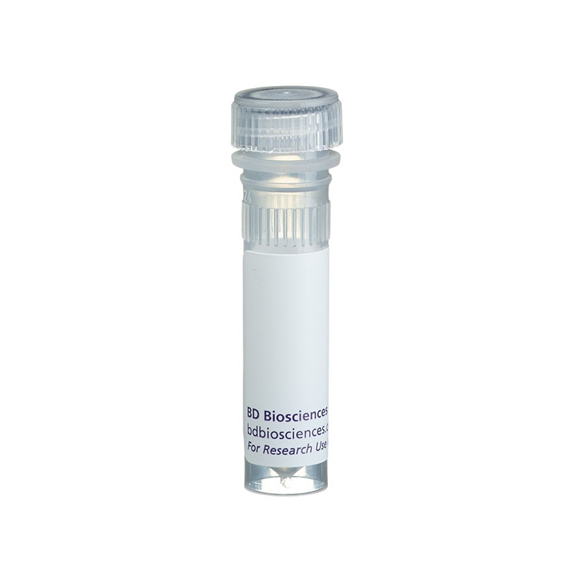
BD Pharmingen™ Purified NA/LE Mouse Anti-Human TNF
Clone MAb1 (RUO)


Regulatory Status Legend
Any use of products other than the permitted use without the express written authorization of Becton, Dickinson and Company is strictly prohibited.
Preparation And Storage
Recommended Assay Procedures
ELISA: Purified Mouse Anti-Human TNF antibody (Clone MAb1, Cat. No. 551220) is useful as a capture antibody for a sandwich ELISA for measuring human TNF. The purified MAb1 antibody can be paired with Biotin Mouse Anti-Human TNF mAb ( Clone MAb11, Cat. No. 554511) as the detection antibody, with recombinant human TNF protein (Cat. No. 554618) as the standard. For complex biological samples, such as serum or plasma, investigators are encouraged to use the BD OptEIA™ Human TNF ELISA Set (Cat. No. 555212) or BD OptEIA™ Human TNF ELISA Kit II (Cat. No. 550610).
Product Notices
- Since applications vary, each investigator should titrate the reagent to obtain optimal results.
- An isotype control should be used at the same concentration as the antibody of interest.
- Please refer to www.bdbiosciences.com/us/s/resources for technical protocols.
Companion Products


The MAb1 antibody reacts with human tumor necrosis factor (TNF, aka TNF-α) protein. TNF is a multifunctional cytokine involved in a variety of immune and inflammatory responses including hemorrhagic necrosis of tumors, septic shock, fever and autoimmune diseases. TNF can regulate the growth and differentiation of many different cell types. It is produced by different activated cell types including macrophages, T lymphocytes, NK cells, dendritic cells, endothelial cells, peripheral blood leukocytes, osteoblasts, astrocytes, mast cells, Kupffer cells, smooth muscle cells, fibroblasts and certain tumor cells. TNF exists in two biologically active forms, i.e., transmembrane and soluble forms. Upon activation, cells express transmembrane TNF glycoproteins that associate as homotrimeric complexes. After enzymatic cleavage, the extracellular region of membrane TNF sheds as a soluble homotrimer. The biologically active form of TNF has been reported to be a trimer.
Development References (3)
-
Danis VA, Franic GM, Rathjen DA, Brooks PM. Effects of granulocyte-macrophage colony-stimulating factor (GM-CSF), IL-2, interferon-gamma (IFN-gamma), tumour necrosis factor-alpha (TNF-alpha) and IL-6 on the production of immunoreactive IL-1 and TNF-alpha by human monocytes. Clin Exp Immunol. 1991; 85(1):143-150. (Clone-specific: ELISA). View Reference
-
Hogan MM, Vogel SN. Production of tumor necrosis factor by rIFN-gamma-primed C3H/HeJ (Lpsd) macrophages requires the presence of lipid A-associated proteins. J Immunol. 1988; 141(12):4196-4202. (Methodology). View Reference
-
Rathjen DA, Cowan K, Furphy LJ, Aston R. Antigenic structure of human tumour necrosis factor: recognition of distinct regions of TNF alpha by different tumour cell receptors. Mol Immunol. 1991; 28(1-2):79-86. (Clone-specific: ELISA). View Reference
Please refer to Support Documents for Quality Certificates
Global - Refer to manufacturer's instructions for use and related User Manuals and Technical data sheets before using this products as described
Comparisons, where applicable, are made against older BD Technology, manual methods or are general performance claims. Comparisons are not made against non-BD technologies, unless otherwise noted.
For Research Use Only. Not for use in diagnostic or therapeutic procedures.