-
Reagents
- Flow Cytometry Reagents
-
Western Blotting and Molecular Reagents
- Immunoassay Reagents
-
Single-Cell Multiomics Reagents
- BD® OMICS-Guard Sample Preservation Buffer
- BD® AbSeq Assay
- BD® Single-Cell Multiplexing Kit
- BD Rhapsody™ ATAC-Seq Assays
- BD Rhapsody™ Whole Transcriptome Analysis (WTA) Amplification Kit
- BD Rhapsody™ TCR/BCR Next Multiomic Assays
- BD Rhapsody™ Targeted mRNA Kits
- BD Rhapsody™ Accessory Kits
- BD® OMICS-One Protein Panels
- BD OMICS-One™ WTA Next Assay
-
Functional Assays
-
Microscopy and Imaging Reagents
-
Cell Preparation and Separation Reagents
Old Browser
This page has been recently translated and is available in French now.
Looks like you're visiting us from {countryName}.
Would you like to stay on the current location site or be switched to your location?
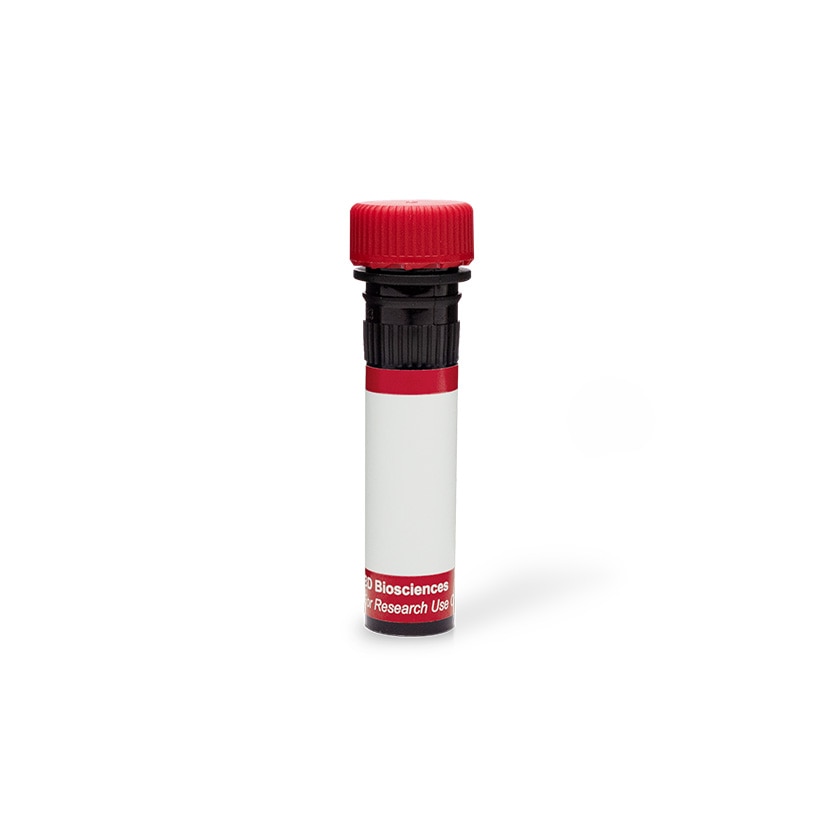
BD Pharmingen™ Alexa Fluor® 647 Mouse anti-SSEA-4
Clone MC813-70 (RUO)

Immunofluorescent staining of human ES cell line. H9 cells (WiCell, Madison, WI) were grown on mouse epithelial feeder cells in a BD Falcon™ 96-well Imaging Plate (Cat. No. 353219). After overnight culture, the cells were fixed and stained with Alexa Fluor® 647 Mouse anti-SSEA-4 monoclonal antibody (pseudo-colored green) and counter-stained with Hoechst 33342 (pseudo-colored blue) according to the Recommended Assay Procedure. The images were captured on a BD Pathway™ 435 Bioimager System with a 10x objective and merged using BD Attovision™ software. This antibody also stained human EC NCCIT (ATCC, Cat. No. CRL-2073), 2102Ep (Andrews et al, 1980), and NTERA-2 cl.D1 (ATCC, Cat. No. CRL-1973), but not mouse EC F9 (ATCC, Cat. No. CRL-1720). If permeabilization is required for staining additional markers, this antibody is compatible with BD Perm/Wash™ Buffer (Cat. No. 554723), but not Triton™ X-100 or methanol permeabilization (see Recommended Assay Procedure).


Immunofluorescent staining of human ES cell line. H9 cells (WiCell, Madison, WI) were grown on mouse epithelial feeder cells in a BD Falcon™ 96-well Imaging Plate (Cat. No. 353219). After overnight culture, the cells were fixed and stained with Alexa Fluor® 647 Mouse anti-SSEA-4 monoclonal antibody (pseudo-colored green) and counter-stained with Hoechst 33342 (pseudo-colored blue) according to the Recommended Assay Procedure. The images were captured on a BD Pathway™ 435 Bioimager System with a 10x objective and merged using BD Attovision™ software. This antibody also stained human EC NCCIT (ATCC, Cat. No. CRL-2073), 2102Ep (Andrews et al, 1980), and NTERA-2 cl.D1 (ATCC, Cat. No. CRL-1973), but not mouse EC F9 (ATCC, Cat. No. CRL-1720). If permeabilization is required for staining additional markers, this antibody is compatible with BD Perm/Wash™ Buffer (Cat. No. 554723), but not Triton™ X-100 or methanol permeabilization (see Recommended Assay Procedure).

Immunofluorescent staining of human ES cell line. H9 cells (WiCell, Madison, WI) were grown on mouse epithelial feeder cells in a BD Falcon™ 96-well Imaging Plate (Cat. No. 353219). After overnight culture, the cells were fixed and stained with Alexa Fluor® 647 Mouse anti-SSEA-4 monoclonal antibody (pseudo-colored green) and counter-stained with Hoechst 33342 (pseudo-colored blue) according to the Recommended Assay Procedure. The images were captured on a BD Pathway™ 435 Bioimager System with a 10x objective and merged using BD Attovision™ software. This antibody also stained human EC NCCIT (ATCC, Cat. No. CRL-2073), 2102Ep (Andrews et al, 1980), and NTERA-2 cl.D1 (ATCC, Cat. No. CRL-1973), but not mouse EC F9 (ATCC, Cat. No. CRL-1720). If permeabilization is required for staining additional markers, this antibody is compatible with BD Perm/Wash™ Buffer (Cat. No. 554723), but not Triton™ X-100 or methanol permeabilization (see Recommended Assay Procedure).


Regulatory Status Legend
Any use of products other than the permitted use without the express written authorization of Becton, Dickinson and Company is strictly prohibited.
Preparation And Storage
Recommended Assay Procedures
1. Seed the cells in appropriate culture medium at an appropriate cell density in a BD Falcon™ 96-well Imaging Plate (Cat. No. 353219), and
culture overnight to 48 hours.
2. Remove the culture medium from the wells, wash (one to two times) with 100 μl of 1× PBS.
3. Fix the cells by adding 100 µl of fresh 3.7% Formaldehyde in PBS or BD Cytofix™ fixation buffer (Cat. No. 554655) to each well and incubating for 10 minutes at room temperature (RT).
4. Remove the fixative from the wells, and wash the wells (one to two times) with 100 μl of 1× PBS.
5. Permeabilize the cells using either cold methanol (a), Triton™ X-100 (b) or Saponin (c):
a. Add 100 µl of -20°C 90% methanol or -20°C BD™ Phosflow Perm Buffer III (Cat. No. 558050) to each well and incubate for 5 minutes at RT.
b. Add 100 µl of 0.1% Triton™ X-100 to each well and incubate for 5 minutes at RT.
Triton is a trademark of The Dow Chemical Company.
c. Add 100 µl of 1× Perm/Wash buffer (Cat. No. 554723) to each well and incubate for 15 to 30 minutes at RT. Continue to use 1× Perm/Wash buffer for all subsequent wash and dilutions steps.
6. Remove the permeabilization buffer from the wells, and wash one to two times with 100 μl of appropriate buffer (either 1× PBS or 1× Perm/Wash buffer, see step 5.c.).
7. Optional blocking step: Remove the wash buffers and block the cells by adding 100 µl of blocking buffer BD Pharmingen™ Stain Buffer (FBS) (Cat. No. 554656) or 3% FBS in appropriate dilution buffer to each well and incubating for 15 to 30 minutes at RT.
8. Dilute the antibody to its optimal working concentration in appropriate dilution buffer. Titrate purified (unconjugated) antibodies and second-step reagents to determine the optimal concentration. If using a Bioimaging Certified antibody conjugate, dilute it 1:10.
9. Add 50 µl of diluted antibody per well and incubate for 60 minutes at RT. Incubate in the dark if using fluorescently labeled antibodies.
10. Remove the antibody, and wash the wells three times with 100 μl of wash buffer. An optional detergent wash (100 μl of 0.05% Tween in 1× PBS) can be included prior to the regular wash steps.
11. If the antibody being used is fluorescently labeled then move to step 12. Otherwise, if using a purified unlabeled antibody, repeat steps 8 to 10 with a fluorescently labeled second-step reagent to detect the purified antibody.
12. After the final wash, counter-stain the nuclei by adding 100 μl of a 2 μg/ml solution of Hoechst 33342 (eg, Sigma-Aldrich Cat. No. B2261) in 1× PBS to each well at least 15 minutes before imaging.
13. View and analyze the cells on an appropriate imaging instrument. Recommended filters for the BD Pathway™ instruments are:
Instrument Excitation Emission Dichroic
BD Pathway 855 620/60 700/75 660 LP
BD Pathway 435 628/40 690/40 FF660
Product Notices
- Please refer to www.bdbiosciences.com/us/s/resources for technical protocols.
- This reagent has been pre-diluted for use at the recommended Volume per Test when following the Recommended Assay Procedure. A Test is typically ~10,000 cells cultured in a well of a 96-well imaging plate.
- Caution: Sodium azide yields highly toxic hydrazoic acid under acidic conditions. Dilute azide compounds in running water before discarding to avoid accumulation of potentially explosive deposits in plumbing.
- Source of all serum proteins is from USDA inspected abattoirs located in the United States.
- Alexa Fluor® is a registered trademark of Molecular Probes, Inc., Eugene, OR.
- The Alexa Fluor®, Pacific Blue™, and Cascade Blue® dye antibody conjugates in this product are sold under license from Molecular Probes, Inc. for research use only, excluding use in combination with microarrays, or as analyte specific reagents. The Alexa Fluor® dyes (except for Alexa Fluor® 430), Pacific Blue™ dye, and Cascade Blue® dye are covered by pending and issued patents.
- Alexa Fluor® 647 fluorochrome emission is collected at the same instrument settings as for allophycocyanin (APC).
Companion Products


The MC813-70 monoclonal antibody reacts with Stage-Specific Embryonic Antigen-4 (SSEA-4), a carbohydrate epitope on the major ganglioside, but not the neutral glycolipid, of human teratocarcinoma cells. As its name implies, the expression of SSEA-4 is stage-specific and can be used to characterize embryonic cells and monitor their differentiation. However, its expression pattern differs in the human and mouse. In the human, SSEA-4 is found on teratocarcinoma (embryonal carcinoma or EC), embryonic inner cell mass (ICM), embryonic stem (ES) cells, and the K562 erythromyeloid leukeumia cell line. As human stem cells undergo differentiation, SSEA-4 expression is lost. In the mouse, SSEA-4 is found on oocytes and early cleavage-stage embryos, and primitive ectoderm, but not on EC, ICM, or ES cells. In some cases, SSEA-4 expression appears upon differentiation of mouse EC or ES cells.

Development References (7)
-
Andrews PW, Bronson DL, Benham F, Strickland S, Knowles BB. A comparative study of eight cell lines derived from human testicular teratocarcinoma. Int J Cancer. 1980; 26(3):269-280. (Methodology). View Reference
-
Draper JS, Pigott C, Thomson JA, Andrews PW. Surface antigens of human embryonic stem cells: changes upon differentiation in culture. J Anat. 2002; 200:249-258. (Clone-specific: Flow cytometry). View Reference
-
Henderson JK, Draper JS, Baillie HS, et al. Preimplantation human embryos and embryonic stem cells show comparable expression of stage-specific embryonic antigens. Stem Cells. 2002; 20:329-337. (Clone-specific: Flow cytometry, Immunocytochemistry (cytospins)). View Reference
-
Josephson R, Ording CJ, Liu Y, et al. Qualification of embryonal carcinoma 2102Ep as a reference for human embryonic stem cell research. Stem Cells. 2007; 25:437-446. (Clone-specific: Flow cytometry, Immunofluorescence). View Reference
-
Kannagi R, Cochran NA, Ishigami F, et al. Stage-specific embryonic antigens (SSEA-3 and -4) are epitopes of a unique globo-series ganglioside isolated from human teratocarcinoma cells. EMBO J. 1983; 2(12):2355-2361. (Immunogen: Immunofluorescence, Radioimmunoassay). View Reference
-
Son YS, Park JH, Kang YK, et al. Heat shock 70-kDa protein 8 isoform 1 is expressed on the surface of human embryonic stem cells and downregulated upon differentiation. Stem Cells. 2005; 23:1502-1513. (Clone-specific: Flow cytometry, Immunocytochemistry (cytospins)). View Reference
-
Thomson JA, Itskovitz-Eldor J, Shapiro SS, et al. Embryonic stem cell lines derived from human blastocysts. Science. 1998; 282:1145-1147. (Clone-specific: Immunocytochemistry (cytospins)). View Reference
Please refer to Support Documents for Quality Certificates
Global - Refer to manufacturer's instructions for use and related User Manuals and Technical data sheets before using this products as described
Comparisons, where applicable, are made against older BD Technology, manual methods or are general performance claims. Comparisons are not made against non-BD technologies, unless otherwise noted.
For Research Use Only. Not for use in diagnostic or therapeutic procedures.