





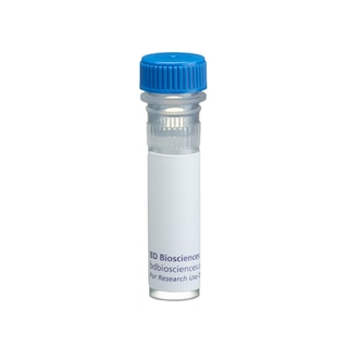
BD Pharmingen™ Purified Mouse anti-SSEA-1
Clone MC480 (RUO)


Western Blot analysis of SSEA-1 in mouse ES cell line. Lysate from ES-E14TG2a cells (ATCC CRL-1821) was probed with Purified Mouse anti-SSEA-1 monoclonal antibody (Cat. No. 560079) at titrations of 1.0 (lane 1), 0.5 (lane 2), and 0.25 µg/ml (lane 3), followed by HRP Goat Anti-Mouse Ig (Cat. No. 554002). High-molecular-weight molecules bearing the SSEA-1 epitope are identified above 200 kDa.

Immunofluorescent staining of mouse ES cell line. ES-E14TG2a cells were cultured, fixed, and stained with Purified Mouse anti-SSEA-1 monoclonal antibody (pseudo-colored green) according to the Recommended Assay Procedure. The second-step reagent was Alexa Fluor® 647 goat anti-mouse Ig (Invitrogen) and counter-staining was with Hoechst 33342 (pseudo-colored blue). The images were captured on a BD Pathway™ 435 Cell Analyzer using a 20X objective and merged using BD Attovision™ software.

Western Blot analysis of SSEA-1 in mouse ES cell line. Lysate from ES-E14TG2a cells (ATCC CRL-1821) was probed with Purified Mouse anti-SSEA-1 monoclonal antibody (Cat. No. 560079) at titrations of 1.0 (lane 1), 0.5 (lane 2), and 0.25 µg/ml (lane 3), followed by HRP Goat Anti-Mouse Ig (Cat. No. 554002). High-molecular-weight molecules bearing the SSEA-1 epitope are identified above 200 kDa.
Immunofluorescent staining of mouse ES cell line. ES-E14TG2a cells were cultured, fixed, and stained with Purified Mouse anti-SSEA-1 monoclonal antibody (pseudo-colored green) according to the Recommended Assay Procedure. The second-step reagent was Alexa Fluor® 647 goat anti-mouse Ig (Invitrogen) and counter-staining was with Hoechst 33342 (pseudo-colored blue). The images were captured on a BD Pathway™ 435 Cell Analyzer using a 20X objective and merged using BD Attovision™ software.


Preparation And Storage


Recommended Assay Procedures
Bioimaging
1. Seed the cells in appropriate culture medium at an appropriate cell density in a Falcon™ 96-well Imaging Plate (Cat. No. 353219), and
culture overnight to 48 hours.
2. Remove the culture medium from the wells, wash the wells twice with 100 μl of 1× PBS, and fix the cells by adding 100 µl of fresh 3.7% Formaldehyde in PBS or BD Cytofix™ fixation buffer (Cat. No. 554655) to each well and incubating for 10 minutes at room temperature (RT).
3. Remove the fixative from the wells, and wash the wells twice with 100 μl of 1× PBS.
4. Dilute the antibody in 1× PBS, and stain the cells by adding 50 µl of the diluted antibody to each well and incubating for 1 hour at RT.
5. Remove the diluted antibody, and wash the wells three times with 100 μl of 1× PBS.
6. Remove the PBS, dilute the second-step reagent in 1× PBS, and stain the cells by adding 50 µl of the diluted second-step reagent to each well and incubating for 1 hour at RT.
7. Remove the diluted second-step reagent, and wash the wells twice with 100 μl of 1× PBS.
8. Remove the PBS, and counter-stain the nuclei by adding 100 μl of a 2 μg/ml solution of Hoechst 33342 (eg, Sigma-Aldrich Cat. No. B2261) in 1× PBS to each well at least 15 minutes before imaging.
9. View and analyze the cells on an appropriate imaging instrument.
Product Notices
- Since applications vary, each investigator should titrate the reagent to obtain optimal results.
- An isotype control should be used at the same concentration as the antibody of interest.
- Caution: Sodium azide yields highly toxic hydrazoic acid under acidic conditions. Dilute azide compounds in running water before discarding to avoid accumulation of potentially explosive deposits in plumbing.
- Sodium azide is a reversible inhibitor of oxidative metabolism; therefore, antibody preparations containing this preservative agent must not be used in cell cultures nor injected into animals. Sodium azide may be removed by washing stained cells or plate-bound antibody or dialyzing soluble antibody in sodium azide-free buffer. Since endotoxin may also affect the results of functional studies, we recommend the NA/LE (No Azide/Low Endotoxin) antibody format, if available, for in vitro and in vivo use.
- This antibody has been developed and certified for the bioimaging application. However, a routine bioimaging test is not performed on every lot. Researchers are encouraged to titrate the reagent for optimal performance.
- Please refer to http://regdocs.bd.com to access safety data sheets (SDS).
- Species cross-reactivity detected in product development may not have been confirmed on every format and/or application.
- Please refer to www.bdbiosciences.com/us/s/resources for technical protocols.
Companion Products

The MC480 monoclonal antibody reacts with Stage-Specific Embryonic Antigen-1 (SSEA-1), which is a terminal carbohydrate epitope (3-fucosyl-N-acetyllactosamine or 3-FAL) on glycoproteins and lactose-series glycolipids. SSEA-1 is related to Lewis blood group antigens and is found in a variety of embryonic as well as adult tissues and cancers. As its name implies, the expression of SSEA-1 is stage-specific and can be used to characterize embryonic cells and monitor their differentiation. However, its expression pattern differs between human and mice. In the human, SSEA-1 is not found on embryonic stem (ES) cells, embryonic inner cell mass (ICM), or teratocarcinoma (embryonal carcinoma or EC) cells. As human EC and ES cells undergo differentiation, SSEA-1 expression is upregulated. In the adult, the same epitope is expressed as CD15 on granulocytes and monocytes, but not lymphocytes or dendritic cells. In the mouse, SSEA-1 is found on EC, ES, primordial germ cells, 8-cell to blastocyst embryos, ICM, and subpopulations of cells in the adult central nervous system, including stem cells. In contrast to human SSEA-1 expression, it is reduced when mouse EC and ES cells undergo differentiation.
Development References (7)
-
Capela A, Temple S. LeX/ssea-1 is expressed by adult mouse CNS stem cells, identifying them as nonependymal. Neuron. 2002; 35:865-875. (Biology).
-
Childs RA, Pennington J, Uemura K, et al. High-molecular-weight glycoproteins are the major carriers of the carbohydrate differentiation antigens I, i and SSEA-1 of mouse teratocarcinoma cells. Biochem J. 1983; 215:491-503. (Clone-specific: Immunofluorescence, Western blot).
-
Draper JS, Pigott C, Thomson JA, Andrews PW. Surface antigens of human embryonic stem cells: changes upon differentiation in culture. J Anat. 2002; 200:249-258. (Clone-specific: Flow cytometry). View Reference
-
Henderson JK, Draper JS, Baillie HS, et al. Preimplantation human embryos and embryonic stem cells show comparable expression of stage-specific embryonic antigens. Stem Cells. 2002; 20:329-337. (Clone-specific: Flow cytometry, Immunofluorescence). View Reference
-
Kannagi R, Nudelman E, Levery SB, Hakomori S. A series of human erythrocyte glycosphingolipids reacting to the monoclonal antibody directed to a developmentally regulated antigen, SSEA-1. J Biol Chem. 1982; 257(24):14865-14874. (Clone-specific).
-
Solter D, Knowles BB. Monoclonal antibody defining a stage-specific mouse embryonic antigen (SSEA-1). Proc Natl Acad Sci U S A. 1978; 75(11):5565-5569. (Immunogen: Cytotoxicity, Radioimmunoassay). View Reference
-
Thomson JA, Itskovitz-Eldor J, Shapiro SS, et al. Embryonic stem cell lines derived from human blastocysts. Science. 1998; 282:1145-1147. (Clone-specific: Immunocytochemistry (cytospins)). View Reference
Please refer to Support Documents for Quality Certificates
Global - Refer to manufacturer's instructions for use and related User Manuals and Technical data sheets before using this products as described
Comparisons, where applicable, are made against older BD Technology, manual methods or are general performance claims. Comparisons are not made against non-BD technologies, unless otherwise noted.
For Research Use Only. Not for use in diagnostic or therapeutic procedures.