The site is undergoing maintenance on
Thursday, September 10, 5:00 pm through Friday, September 11, 1:00 am (CEST), 2026.
If you encounter problems while ordering, please contact us at BDBCustomerService@bd.com
See More
The site is undergoing maintenance on
Thursday, September 10, 5:00 pm through Friday, September 11, 1:00 am (CEST), 2026.
If you encounter problems while ordering, please contact us at BDBCustomerService@bd.com
See MoreThe site is undergoing maintenance on
Thursday, September 10, 5:00 pm through Friday, September 11, 1:00 am (CEST), 2026
.If you encounter problems while ordering, please contact us at BDBCustomerService@bd.com
This page has been recently translated and is available in French now.
Looks like you're visiting us from {countryName}.
Would you like to stay on the current location site or be switched to your location?
Analyses of mTOR (pS2448) expression in stimulated human peripheral blood B cells.
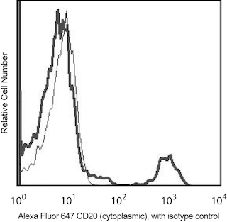
Left Panel: Flow cytometric analysis of mTOR (pS2448) expression. Human B lymphocytes were prepared from peripheral blood mononuclear cells (PBMC) by negative selection using the BD IMag™ Human B Lymphocyte Enrichment Set - DM (Cat. No. 558007). The B cells were serum starved overnight in culture and were either not stimulated (dashed line histogram) or were stimulated with Type C CpG oligonucleotide (1 μM, 4 hrs; InvivoGen, Cat. No. TLRL-2395). The cells were fixed with BD Cytofix™ Fixation Buffer (Cat. No. 554655; 10 minutes at 37°C) and then permeabilized by adding BD Phosflow™ Perm Buffer III (Cat. No. 558050; 30 minutes on ice). The cells were washed twice with BD Pharmingen™ Stain Buffer (Cat. No. 554656) and then stained with BD Phosflow™ PE Mouse Anti-mTOR (pS2448) (Cat. No. 563489) and Alexa Fluor® 647 Mouse Anti-Human CD20 (Cat. No. 558054) antibodies using the BD Biosciences Protocol for Intracellular Staining. The fluorescence histograms were derived from CD20 positive-gated events with the forward and side light-scatter characteristics of intact lymphocytes. Flow cytometric analysis was performed using a BD FACSCanto™ II Flow Cytometer.
Right Panel: Western blot analysis of mTOR (pS2448) expression. Aliquots of the unstimulated (lane 1) or CPG-stimulated (lane 2) B cells described above were made into lysates. The lysates were electrophoresed, transferred to membranes and blotted using Purified Mouse Anti-mTOR (pS2448) antibody (Clone O21-404; 2 μg/mL). Phosphorylated mTOR (pS2448) was identified as ~240-245 kDa band.
Analyses of mTOR (pS2448) expression in stimulated human peripheral blood B cells.
Left Panel: Flow cytometric analysis of mTOR (pS2448) expression. Human B lymphocytes were prepared from peripheral blood mononuclear cells (PBMC) by negative selection using the BD IMag™ Human B Lymphocyte Enrichment Set - DM (Cat. No. 558007). The B cells were serum starved overnight in culture and were either not stimulated (dashed line histogram) or were stimulated with Type C CpG oligonucleotide (1 μM, 4 hrs; InvivoGen, Cat. No. TLRL-2395). The cells were fixed with BD Cytofix™ Fixation Buffer (Cat. No. 554655; 10 minutes at 37°C) and then permeabilized by adding BD Phosflow™ Perm Buffer III (Cat. No. 558050; 30 minutes on ice). The cells were washed twice with BD Pharmingen™ Stain Buffer (Cat. No. 554656) and then stained with BD Phosflow™ PE Mouse Anti-mTOR (pS2448) (Cat. No. 563489) and Alexa Fluor® 647 Mouse Anti-Human CD20 (Cat. No. 558054) antibodies using the BD Biosciences Protocol for Intracellular Staining. The fluorescence histograms were derived from CD20 positive-gated events with the forward and side light-scatter characteristics of intact lymphocytes. Flow cytometric analysis was performed using a BD FACSCanto™ II Flow Cytometer.
Right Panel: Western blot analysis of mTOR (pS2448) expression. Aliquots of the unstimulated (lane 1) or CPG-stimulated (lane 2) B cells described above were made into lysates. The lysates were electrophoresed, transferred to membranes and blotted using Purified Mouse Anti-mTOR (pS2448) antibody (Clone O21-404; 2 μg/mL). Phosphorylated mTOR (pS2448) was identified as ~240-245 kDa band.
Analyses of mTOR (pS2448) expression in stimulated human peripheral blood B cells.
Left Panel: Flow cytometric analysis of mTOR (pS2448) expression. Human B lymphocytes were prepared from peripheral blood mononuclear cells (PBMC) by negative selection using the BD IMag™ Human B Lymphocyte Enrichment Set - DM (Cat. No. 558007). The B cells were serum starved overnight in culture and were either not stimulated (dashed line histogram) or were stimulated with Type C CpG oligonucleotide (1 μM, 4 hrs; InvivoGen, Cat. No. TLRL-2395). The cells were fixed with BD Cytofix™ Fixation Buffer (Cat. No. 554655; 10 minutes at 37°C) and then permeabilized by adding BD Phosflow™ Perm Buffer III (Cat. No. 558050; 30 minutes on ice). The cells were washed twice with BD Pharmingen™ Stain Buffer (Cat. No. 554656) and then stained with BD Phosflow™ PE Mouse Anti-mTOR (pS2448) (Cat. No. 563489) and Alexa Fluor® 647 Mouse Anti-Human CD20 (Cat. No. 558054) antibodies using the BD Biosciences Protocol for Intracellular Staining. The fluorescence histograms were derived from CD20 positive-gated events with the forward and side light-scatter characteristics of intact lymphocytes. Flow cytometric analysis was performed using a BD FACSCanto™ II Flow Cytometer.
Right Panel: Western blot analysis of mTOR (pS2448) expression. Aliquots of the unstimulated (lane 1) or CPG-stimulated (lane 2) B cells described above were made into lysates. The lysates were electrophoresed, transferred to membranes and blotted using Purified Mouse Anti-mTOR (pS2448) antibody (Clone O21-404; 2 μg/mL). Phosphorylated mTOR (pS2448) was identified as ~240-245 kDa band.
Analyses of mTOR (pS2448) expression in stimulated human peripheral blood B cells.
Left Panel: Flow cytometric analysis of mTOR (pS2448) expression. Human B lymphocytes were prepared from peripheral blood mononuclear cells (PBMC) by negative selection using the BD IMag™ Human B Lymphocyte Enrichment Set - DM (Cat. No. 558007). The B cells were serum starved overnight in culture and were either not stimulated (dashed line histogram) or were stimulated with Type C CpG oligonucleotide (1 μM, 4 hrs; InvivoGen, Cat. No. TLRL-2395). The cells were fixed with BD Cytofix™ Fixation Buffer (Cat. No. 554655; 10 minutes at 37°C) and then permeabilized by adding BD Phosflow™ Perm Buffer III (Cat. No. 558050; 30 minutes on ice). The cells were washed twice with BD Pharmingen™ Stain Buffer (Cat. No. 554656) and then stained with BD Phosflow™ PE Mouse Anti-mTOR (pS2448) (Cat. No. 563489) and Alexa Fluor® 647 Mouse Anti-Human CD20 (Cat. No. 558054) antibodies using the BD Biosciences Protocol for Intracellular Staining. The fluorescence histograms were derived from CD20 positive-gated events with the forward and side light-scatter characteristics of intact lymphocytes. Flow cytometric analysis was performed using a BD FACSCanto™ II Flow Cytometer.
Right Panel: Western blot analysis of mTOR (pS2448) expression. Aliquots of the unstimulated (lane 1) or CPG-stimulated (lane 2) B cells described above were made into lysates. The lysates were electrophoresed, transferred to membranes and blotted using Purified Mouse Anti-mTOR (pS2448) antibody (Clone O21-404; 2 μg/mL). Phosphorylated mTOR (pS2448) was identified as ~240-245 kDa band.
Analyses of mTOR (pS2448) expression in stimulated human peripheral blood B cells.
Left Panel: Flow cytometric analysis of mTOR (pS2448) expression. Human B lymphocytes were prepared from peripheral blood mononuclear cells (PBMC) by negative selection using the BD IMag™ Human B Lymphocyte Enrichment Set - DM (Cat. No. 558007). The B cells were serum starved overnight in culture and were either not stimulated (dashed line histogram) or were stimulated with Type C CpG oligonucleotide (1 μM, 4 hrs; InvivoGen, Cat. No. TLRL-2395). The cells were fixed with BD Cytofix™ Fixation Buffer (Cat. No. 554655; 10 minutes at 37°C) and then permeabilized by adding BD Phosflow™ Perm Buffer III (Cat. No. 558050; 30 minutes on ice). The cells were washed twice with BD Pharmingen™ Stain Buffer (Cat. No. 554656) and then stained with BD Phosflow™ PE Mouse Anti-mTOR (pS2448) (Cat. No. 563489) and Alexa Fluor® 647 Mouse Anti-Human CD20 (Cat. No. 558054) antibodies using the BD Biosciences Protocol for Intracellular Staining. The fluorescence histograms were derived from CD20 positive-gated events with the forward and side light-scatter characteristics of intact lymphocytes. Flow cytometric analysis was performed using a BD FACSCanto™ II Flow Cytometer.
Right Panel: Western blot analysis of mTOR (pS2448) expression. Aliquots of the unstimulated (lane 1) or CPG-stimulated (lane 2) B cells described above were made into lysates. The lysates were electrophoresed, transferred to membranes and blotted using Purified Mouse Anti-mTOR (pS2448) antibody (Clone O21-404; 2 μg/mL). Phosphorylated mTOR (pS2448) was identified as ~240-245 kDa band.
Analyses of mTOR (pS2448) expression in stimulated human peripheral blood B cells.
Left Panel: Flow cytometric analysis of mTOR (pS2448) expression. Human B lymphocytes were prepared from peripheral blood mononuclear cells (PBMC) by negative selection using the BD IMag™ Human B Lymphocyte Enrichment Set - DM (Cat. No. 558007). The B cells were serum starved overnight in culture and were either not stimulated (dashed line histogram) or were stimulated with Type C CpG oligonucleotide (1 μM, 4 hrs; InvivoGen, Cat. No. TLRL-2395). The cells were fixed with BD Cytofix™ Fixation Buffer (Cat. No. 554655; 10 minutes at 37°C) and then permeabilized by adding BD Phosflow™ Perm Buffer III (Cat. No. 558050; 30 minutes on ice). The cells were washed twice with BD Pharmingen™ Stain Buffer (Cat. No. 554656) and then stained with BD Phosflow™ PE Mouse Anti-mTOR (pS2448) (Cat. No. 563489) and Alexa Fluor® 647 Mouse Anti-Human CD20 (Cat. No. 558054) antibodies using the BD Biosciences Protocol for Intracellular Staining. The fluorescence histograms were derived from CD20 positive-gated events with the forward and side light-scatter characteristics of intact lymphocytes. Flow cytometric analysis was performed using a BD FACSCanto™ II Flow Cytometer.
Right Panel: Western blot analysis of mTOR (pS2448) expression. Aliquots of the unstimulated (lane 1) or CPG-stimulated (lane 2) B cells described above were made into lysates. The lysates were electrophoresed, transferred to membranes and blotted using Purified Mouse Anti-mTOR (pS2448) antibody (Clone O21-404; 2 μg/mL). Phosphorylated mTOR (pS2448) was identified as ~240-245 kDa band.
Any use of products other than the permitted use without the express written authorization of Becton, Dickinson and Company is strictly prohibited.
The O21-404 monoclonal antibody recognizes the human mammalian target of rapmycin,(mTOR) that is phosphorylated at
serine residue 2448, mTOR (pS2448). mTOR belongs to the phosphoinositide-3-kinase (PI3K)-related (PIKK) family of kinases. mTOR is also known as mechanistic target of rapamycin (serine/threonine kinase), FRAP, RAFT1, and RAPT1. mTOR functions as an amino acid and ATP sensor to balance nutrient availability and cell growth. When nutrients are sufficiently available, mTOR is activated by phosphorylation at serine residue 2448 through the PI3 kinase/Akt signaling pathway. Phosphorylated mTOR in turn activates the p70 S6 kinase and contributes to the inactivation 4E-BP1, an elF4E inhibitor. In this way, mTOR participates in the upregulation of cellular protein synthesis, growth, proliferation and survival. mTOR function may be abnormally regulated in tumors.

Please refer to Support Documents for Quality Certificates
Global - Refer to manufacturer's instructions for use and related User Manuals and Technical data sheets before using this products as described
Comparisons, where applicable, are made against older BD Technology, manual methods or are general performance claims. Comparisons are not made against non-BD technologies, unless otherwise noted.
For Research Use Only. Not for use in diagnostic or therapeutic procedures.