





.png)


Preparation And Storage


Recommended Assay Procedures
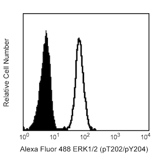
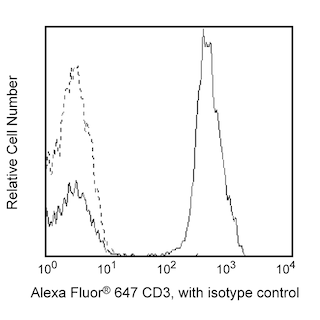
Immunofluorescent staining with the O4-46 Mouse Anti-T-bet monoclonal antibody is compatible with BD Phosflow™ Lyse/Fix Buffer (Cat. No. 558049) and the BD Phosflow™ Permeabilization Buffers: Perm/Wash Buffer I (Cat. No. 557885); Perm Buffer II (Cat. No. 558052); Perm Buffer III (Cat. No. 558050); and Perm Buffer IV (Cat. No. 560746).
Product Notices
- This reagent has been pre-diluted for use at the recommended Volume per Test. We typically use 1 × 10^6 cells in a 100-µl experimental sample (a test).
- Source of all serum proteins is from USDA inspected abattoirs located in the United States.
- An isotype control should be used at the same concentration as the antibody of interest.
- Please observe the following precautions: Absorption of visible light can significantly alter the energy transfer occurring in any tandem fluorochrome conjugate; therefore, we recommend that special precautions be taken (such as wrapping vials, tubes, or racks in aluminum foil) to prevent exposure of conjugated reagents, including cells stained with those reagents, to room illumination.
- Caution: Sodium azide yields highly toxic hydrazoic acid under acidic conditions. Dilute azide compounds in running water before discarding to avoid accumulation of potentially explosive deposits in plumbing.
- For fluorochrome spectra and suitable instrument settings, please refer to our Multicolor Flow Cytometry web page at www.bdbiosciences.com/colors.
- Texas Red is a registered trademark of Molecular Probes, Inc., Eugene, OR.
- CF™ is a trademark of Biotium, Inc.
- When excited by the yellow-green (561-nm) laser, the fluorescence may be brighter than when excited by the blue (488-nm) laser.
- This product is provided under an Agreement between BIOTIUM and BD Biosciences. The manufacture, use, sale, offer for sale, or import of this product is subject to one or more patents or pending applications owned or licensed by Biotium, Inc. This product, and only in the amount purchased by buyer, may be used solely for buyer’s own internal research, in a manner consistent with the accompanying product literature. No other right to use, sell or otherwise transfer (a) this product, or (b) its components is hereby granted expressly, by implication or by estoppel. This product is for research use only. Diagnostic uses require a separate license from Biotium, Inc. For information on purchasing a license to this product including for purposes other than research, contact Biotium, Inc., 3159 Corporate Place, Hayward, CA 94545, Tel: (510) 265-1027. Fax: (510) 265-1352. Email: btinfo@biotium.com.
- Because of the broad absorption spectrum of the tandem fluorochrome, extra care must be taken when using multi-laser cytometers, which may directly excite both PE and CF™594.
- This product may be covered by US Patent No. 7,365,169.
- Limited Use License: The buyer (a) shall not sell or otherwise transfer this product to any third party, (b) shall use this product only for its internal, non-clinical research use, (c) shall not use this product for Commercial Purposes without a commercial license from Harvard (and if the buyer is interested in a commercial license, it should contact Harvard’s Office of Technology Development at 1350 Massachusetts Avenue, Holyoke Center, Suite 727, Cambridge, MA 02138, 617-495-3067), (d) shall use this product in compliance with all applicable laws and regulations, including, without limitation, applicable human health and animal welfare laws and regulations, (e) may transfer information or materials made through the use of this product to a scientific collaborator only if such transfer is not for any Commercial Purpose and such collaborator agrees in writing not to transfer such materials to any third party and to use such transferred materials and/or information solely for internal, non-clinical research and not for Commercial Purposes, (f) acknowledges that this product has not been approved for use in humans by the U.S. Food and Drug Administration or any other regulatory body and may not be used in humans and (g) shall indemnify, defend and hold harmless Becton Dickinson and Company and Harvard from and against all damages, losses, expenses (including reasonable attorneys’ fees), claims, demands, suits and other actions in any way arising from the buyer’s use, storage or disposal of this product.
- Species cross-reactivity detected in product development may not have been confirmed on every format and/or application.
- Alexa Fluor® is a registered trademark of Molecular Probes, Inc., Eugene, OR.
- Cy is a trademark of GE Healthcare.
- Please refer to www.bdbiosciences.com/us/s/resources for technical protocols.
Companion Products


.png?imwidth=320)
The O4-46 monoclonal antibody specifically binds to human and mouse T-bet. T-bet (T-box gene expressed in T cells) is a master regulatory transcription factor that is also known as TBX21 (T-box21) and TBLYM (T-box transcription factor, expressed in lymphocytes). Human (535 amino acids; 58.3 kDa predicted molecular mass) and mouse (530 amino acids; 57.7 kDa) T-bet proteins are encoded by the human TBX21 (chromosome 17) and mouse Tbx21 (chromosome 11) genes. The human and mouse T-bet protein amino acid sequences are 88% homologous. Human and mouse T-bet proteins share a highly conserved (98% homologous amino acid sequences) T-box protein domain that is centrally located and mediates binding to DNA. T-bet is expressed by and activates transcriptional activities within hemotopoietic cells including stem cells, NK and NKT cells and subsets of thymocytes, primed/activated CD4+ T cells, CD8+ T cells and γδ T cells, B cells, and dendritic cells. Interferon-gamma (IFN-γ), interleukin-27 (IL-27), and IL-12 act on peripheral antigen-triggered (TCR-signaling) T cells to increase T-bet expression. With respect to T helper lymphocytes, T-bet directs the differentiation of naïve CD4+ precursor T cells to become Th1-like effector and memory cells. T-bet accomplishes this by activating Th1 genetic programs (including epigenetic modifications) while repressing opposing T helper subset programs. T-bet controls the upregulated expression of the Th1 signature cytokine, IFN-γ, the IL-12Rβ2 subunit and the Runx3 transcription factor and can repress the function of other transcriptional regulators, such as GATA-3 (master regulator of Th2 development) and the expression of other cytokines including IL-2, IL-4 and IL-5.
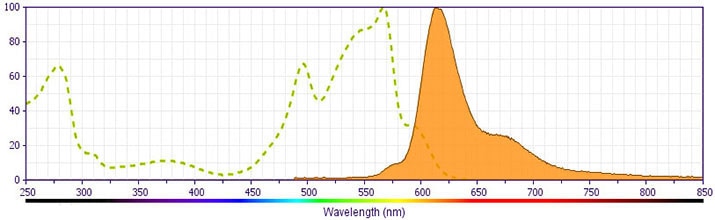
This antibody is conjugated to BD Horizon™ PE-CF594, which has been developed exclusively by BD Biosciences as a better alternative to PE-Texas Red®. PE-CF594 excites and emits at similar wavelengths to PE-Texas Red® yet exhibits improved brightness and spectral characteristics. Due to PE having maximal absorption peaks at 496 nm and 564 nm, PE-CF594 can be excited by the blue (488-nm), green (532-nm) and yellow-green (561-nm) lasers and can be detected with the same filter set as PE-Texas Red® (eg 610/20-nm filter).
Development References (10)
-
Hibbert L, Pflanz S, De Waal Malefyt R, Kastelein RA. IL-27 and IFN-alpha signal via Stat1 and Stat3 and induce T-Bet and IL-12Rbeta2 in naive T cells. J Interferon Cytokine Res. 2003; 23(9):513-522. (Biology). View Reference
-
Hwang ES, Hong JH, Glimcher LH. IL-2 production in developing Th1 cells is regulated by heterodimerization of RelA and T-bet and requires T-bet serine residue 508. J Exp Med. 2005; 202(9):1289-1300. (Biology). View Reference
-
Hwang ES, Szabo SJ, Schwartzberg PL, Glimcher LH. T helper cell fate specified by kinase-mediated interaction of T-bet with GATA-3. Science. 2005; 307(5780):430-433. (Biology). View Reference
-
Lugo-Villarino G, Maldonado-Lopez R, Possemato R, Penaranda C, Glimcher LH. T-bet is required for optimal production of IFN-gamma and antigen-specific T cell activation by dendritic cells. Proc Natl Acad Sci U S A. 2003; 100(13):7749-7754. (Biology). View Reference
-
Peng SL, Szabo SJ, Glimcher LH. T-bet regulates IgG class switching and pathogenic autoantibody production. Proc Natl Acad Sci U S A. 2002; 99(8):5545-5550. (Biology). View Reference
-
Peng SL. The T-box transcription factor T-bet in immunity and autoimmunity. Cell Mol Immunol. 2006; 3(2):87-95. (Biology). View Reference
-
Szabo SJ, Kim ST, Costa GL, Zhang X, Fathman CG, Glimcher LH. A novel transcription factor, T-bet, directs Th1 lineage commitment. Cell. 2000; 100(6):655-669. (Biology). View Reference
-
Takeda A, Hamano S, Yamanaka A, et al. Cutting edge: role of IL-27/WSX-1 signaling for induction of T-bet through activation of STAT1 during initial Th1 commitment. J Immunol. 2003; 170(10):4886-4890. (Biology). View Reference
-
Townsend MJ, Weinmann AS, Matsuda JL, et al. T-bet regulates the terminal maturation and homeostasis of NK and Valpha14i NKT cells. Immunity. 2004; 20(4):477-494. (Biology). View Reference
-
Zhang WX, Yang SY. Cloning and characterization of a new member of the T-box gene family. Genomics. 2000; 70(1):41-48. (Biology). View Reference
Please refer to Support Documents for Quality Certificates
Global - Refer to manufacturer's instructions for use and related User Manuals and Technical data sheets before using this products as described
Comparisons, where applicable, are made against older BD Technology, manual methods or are general performance claims. Comparisons are not made against non-BD technologies, unless otherwise noted.
For Research Use Only. Not for use in diagnostic or therapeutic procedures.
