


Product Details
Description
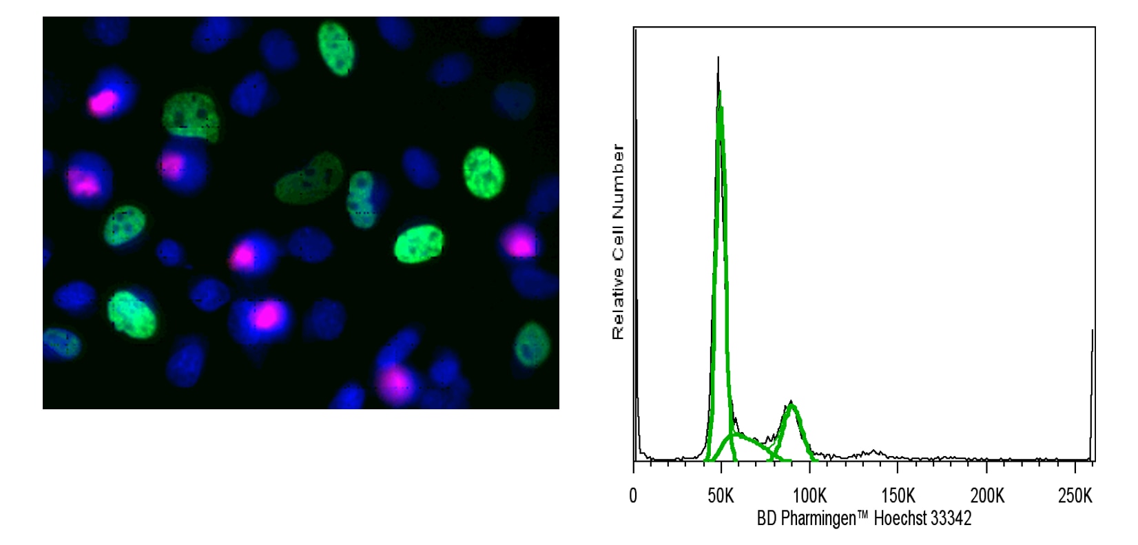
The BD Pharmingen™ Hoechst 33342 Solution is a reagent for the fluorescent staining of DNA and nuclei in live or fixed cells. Hoechst 33342 is a bisbenzimidazole dye with high specificity for binding to double-stranded DNA (preferentially binds to A-T base pairs). This dye is very useful to label double-stranded DNA and thus to visualize nuclei. Hoechst 33342 can be excited at ~355 nm by a UV light source (eg, UV laser beam or a mercury arc-lamp). It emits blue fluorescence light around an emission maximum at 461 nm when bound to DNA. Since the Hoechst 33342 dye is specific for DNA binding, ribonuclease treatment is not needed to avoid nonspecific RNA staining. In addition to its use in fluorescence microscopy and image analysis, Hoechst 33342 is commonly used for flow cytometric applications, such as cell cycle analysis and stem cell side population identification.
Preparation And Storage
Recommended Assay Procedures
Product Notices
- Please refer to www.bdbiosciences.com/us/s/resources for technical protocols.
- This antibody has been developed and certified for the bioimaging application. However, a routine bioimaging test is not performed on every lot. Researchers are encouraged to titrate the reagent for optimal performance.
- Since applications vary, each investigator should titrate the reagent to obtain optimal results.
- Alexa Fluor® is a registered trademark of Molecular Probes, Inc., Eugene, OR.
- FlowJo is a trademark of Tree Star Inc.

Please refer to Support Documents for Quality Certificates
Global - Refer to manufacturer's instructions for use and related User Manuals and Technical data sheets before using this products as described
Comparisons, where applicable, are made against older BD Technology, manual methods or are general performance claims. Comparisons are not made against non-BD technologies, unless otherwise noted.
For Research Use Only. Not for use in diagnostic or therapeutic procedures.