







Preparation And Storage


Recommended Assay Procedures
For optimal and reproducible results, BD Horizon Brilliant Stain Buffer should be used anytime two or more BD Horizon Brilliant dyes are used in the same experiment. Fluorescent dye interactions may cause staining artifacts which may affect data interpretation. The BD Horizon Brilliant Stain Buffer was designed to minimize these interactions. More information can be found in the Technical Data Sheet of the BD Horizon Brilliant Stain Buffer (Cat. No. 563794).
Product Notices
- This reagent has been pre-diluted for use at the recommended Volume per Test. We typically use 1 × 10^6 cells in a 100-µl experimental sample (a test).
- An isotype control should be used at the same concentration as the antibody of interest.
- Source of all serum proteins is from USDA inspected abattoirs located in the United States.
- Caution: Sodium azide yields highly toxic hydrazoic acid under acidic conditions. Dilute azide compounds in running water before discarding to avoid accumulation of potentially explosive deposits in plumbing.
- Species cross-reactivity detected in product development may not have been confirmed on every format and/or application.
- For fluorochrome spectra and suitable instrument settings, please refer to our Multicolor Flow Cytometry web page at www.bdbiosciences.com/colors.
- BD Horizon Brilliant Stain Buffer is covered by one or more of the following US patents: 8,110,673; 8,158,444; 8,575,303; 8,354,239.
- BD Horizon Brilliant Ultraviolet 395 is covered by one or more of the following US patents: 8,158,444; 8,575,303; 8,354,239.
- Please refer to www.bdbiosciences.com/us/s/resources for technical protocols.
Companion Products




The 3G8 monoclonal antibody specifically recognizes CD16a and CD16b, low affinity receptors for the Fc region of IgG. CD16a is ~50-65 kDa type I transmembrane glycoprotein that is encoded by FCGR3A (Fc fragment of IgG receptor IIIa) which belongs to the immunoglobulin superfamily. CD16a is also known as Fc-gamma RIII-alpha (Fc-gamma RIIIa or FcγRIIIA) or FcRIIIa and is expressed on natural killer cells, activated monocytes, macrophages, γδ T cells, immature thymocytes, and mast cells. CD16a binds immune-complexed or aggregated IgG and associates with CD247/TCRζ in NK cells and FcεRIγ chains in phagocytes and mast cells to transduce intracellular signals. CD16a functions in antibody-dependent cellular cytotoxicity (ADCC) and other antibody-dependent responses including phagocytosis, cytokine production or mediator release. CD16b is a ~48 kDa glycophosyl-phosphatidylinositol (GPI)-linked form that is encoded by FCGR3B (Fc fragment of IgG receptor IIIb). CD16b is also known as Fc-gamma RIII-beta (Fc-gamma RIIIb or FcγRIIIB) or FcRIIIb and is expressed on neutrophils and activated eosinophils. The extracellular region of CD16b is highly homologous to CD16a. CD16b also serves as a receptor for the Fc region of IgG and can bind immune-complexed or aggregated IgG and may be involved in neutrophil adhesion.
The 3G8 antibody also crossreacts with a subset of peripheral blood lymphocytes and monocytes, but not granulocytes, of baboon, rhesus, and cynomolgus monkeys. Multicolor analysis reveals that the distribution on lymphocytes is similar to that found in human studies with the majority of CD16-positive lymphocytes being both CD3 and CD20 negative.
This clone also cross-reacts with a subset of peripheral blood lymphocytes and monocytes, but not granulocytes, of baboon and both rhesus and cynomologus macaque monkeys. Multi-color analysis reveals that the distribution on lymphocytes is similar to that found in human studies with the majority of CD16-positive lymphocytes being both CD3 and CD20 negative.
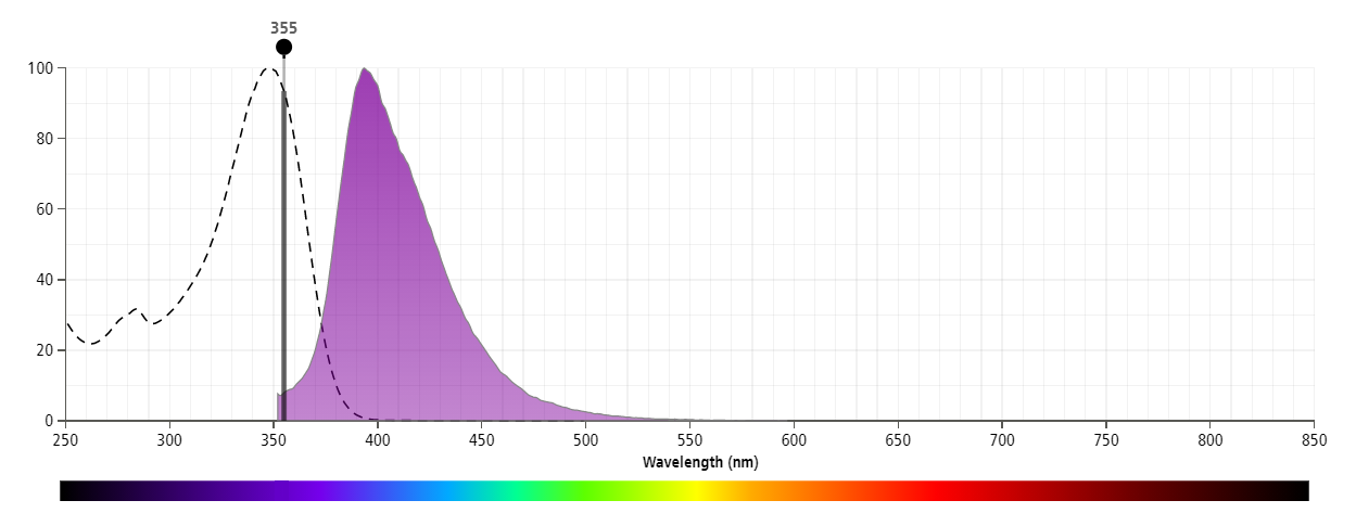
The antibody was conjugated to BD Horizon™ BUV395 which has been exclusively developed by BD Biosciences as an optimal dye for use on a 355 nm laser equipped instrument. With an Ex Max at 348 nm and an Em Max at 395 nm, this dye has virtually no spillover into any other detector. BD Horizon™ BUV395 can be excited with a 355 nm laser and detected with a 379/28 filter.
Development References (8)
-
Fleit HB, Wright SD, Unkeless JC. Human neutrophil Fc gamma receptor distribution and structure. Proc Natl Acad Sci U S A. 1982; 79(10):3275-3279. (Immunogen: Blocking, Immunoprecipitation, Inhibition, Radioimmunoassay). View Reference
-
Knapp W. W. Knapp .. et al., ed. Leucocyte typing IV : white cell differentiation antigens. Oxford New York: Oxford University Press; 1989:1-1182.
-
Perussia B, Trinchieri G, Jackson A, et al. The Fc receptor for IgG on human natural killer cells: phenotypic, functional, and comparative studies with monoclonal antibodies. J Immunol. 1984; 133(1):180-189. (Clone-specific: Flow cytometry, Functional assay, Inhibition). View Reference
-
Schmidt RE. Non-lineage/natural killer section report: new and previously defined clusters. In: Knapp W. W. Knapp .. et al., ed. Leucocyte typing IV : white cell differentiation antigens. Oxford New York: Oxford University Press; 1989:517-542.
-
Stroncek DF, Skubitz KM, Plachta LB, et al. Alloimmune neonatal neutropenia due to an antibody to the neutrophil Fc-gamma receptor III with maternal deficiency of CD16 antigen. Blood. 1991; 77(7):1572-1580. (Clone-specific: Immunofluorescence, Immunoprecipitation). View Reference
-
Vossebeld PJ, Homburg CH, Roos D, Verhoeven AJ. The anti-Fc gamma RIII mAb 3G8 induces neutrophil activation via a cooperative actin of Fc gamma RIIIb and Fc gamma RIIa. Int J Biochem Cell Biol. 1997; 29(3):465-473. (Clone-specific: Activation, Functional assay). View Reference
-
Wirthmueller U, Kurosaki T, Murakami MS, Ravetch JV. Signal transduction by Fc gamma RIII (CD16) is mediated through the gamma chain. J Exp Med. 1992; 175(5):1381-1390. (Clone-specific: Activation, Functional assay). View Reference
-
Zola H. Leukocyte and stromal cell molecules : the CD markers. Hoboken, N.J.: Wiley-Liss; 2007.
Please refer to Support Documents for Quality Certificates
Global - Refer to manufacturer's instructions for use and related User Manuals and Technical data sheets before using this products as described
Comparisons, where applicable, are made against older BD Technology, manual methods or are general performance claims. Comparisons are not made against non-BD technologies, unless otherwise noted.
For Research Use Only. Not for use in diagnostic or therapeutic procedures.
