Old Browser
This page has been recently translated and is available in French now.
Looks like you're visiting us from {countryName}.
Would you like to stay on the current country site or be switched to your country?
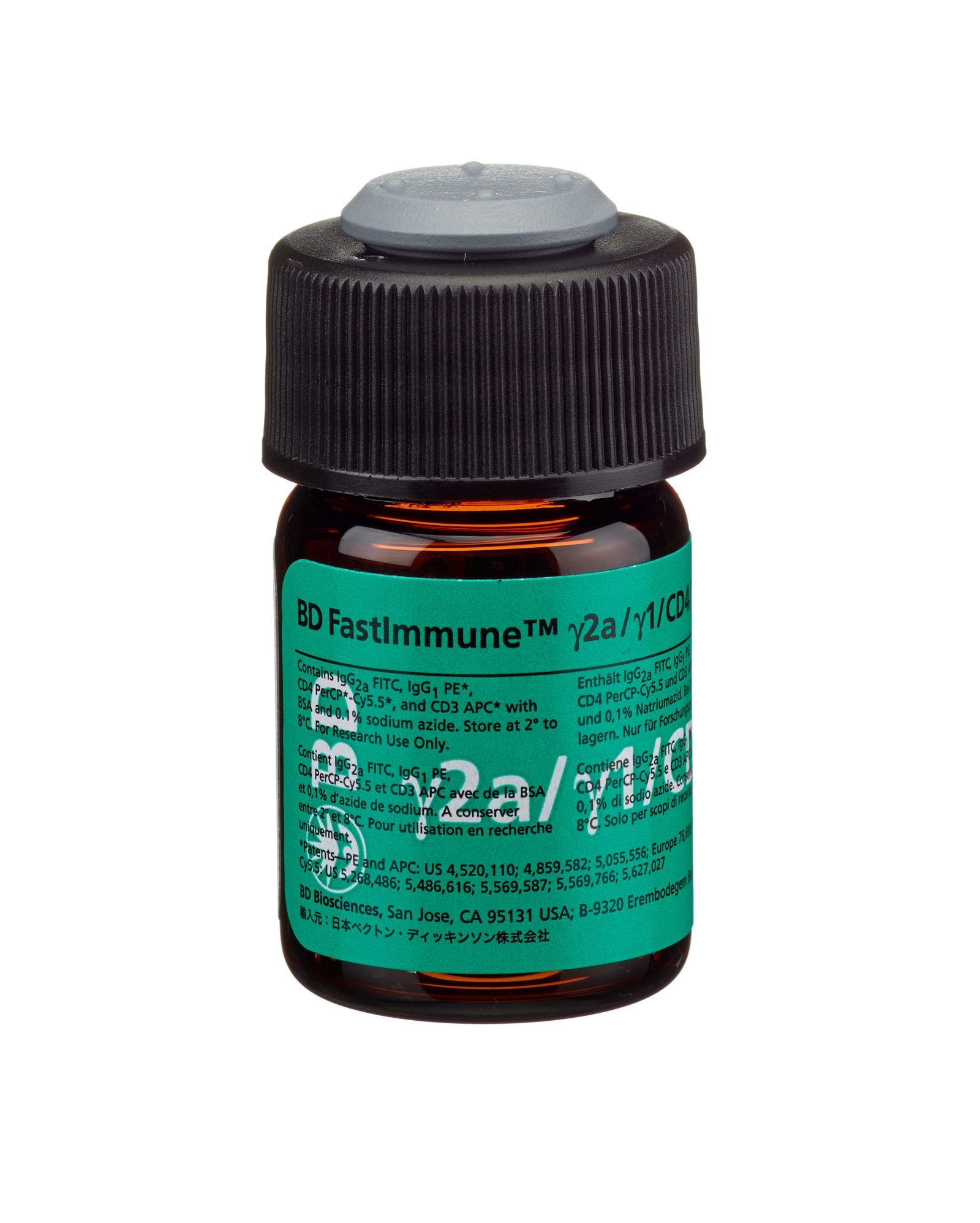
BD FastImmune™ Anti-Human γ2a FITC/γ1 PE/CD4 PerCP-Cy™5.5/CD3 APC
(RUO (GMP))

Anti-Human γ2a FITC/γ1 PE/CD4 PerCP-Cy™5.5/CD3 APC
Regulatory Status Legend
Any use of products other than the permitted use without the express written authorization of Becton, Dickinson and Company is strictly prohibited.
Description
γ2a (IgG2a), clone X39, and γ1 (IgG1), clone X40, are both derived from hybridization of mouse Sp2/0-Ag14 myeloma cells with spleen cells from BALB/c mice immunized with KLH. CD4, clone SK3, is derived from hybridization of mouse NS-1 myeloma cells with spleen cells from BALB/c mice immununized with human peripheral blood T lymphocytes. CD3, clone SK7, is derived from hybridization of mouse NS-1 myeloma cells with spleen cells from BALB/c mice immunized with human thymocytes. Both γ2a (IgG2a) and γ1 (IgG1) react specifically with keyhole limpet hemocyanin (KLH), an antigen not expressed on human cells or human cell lines. The CD4 antibody recognizes an antigen, with a molecular weight of 55-kilodalton (kDa) that is present on T-helper/inducer lymphocytes and monocytes. CD3 reacts with the epsilon chain of the CD3 antigen/T-cell antigen receptor (TCR) complex. This complex is composed of at least six proteins that range in molecular weight from 20 to 30 kd. The antigen recognized by CD3 antibodies is noncovalently associated with either α/β or γ/δ TCR (70 to 90 kd).
Preparation And Storage
Store vials at 2°C–8°C. Conjugated forms should not be frozen. Protect from exposure to light. Each reagent is stable until the expiration date shown on the bottle label when stored as directed.
| Description | Clone | Isotype | EntrezGene ID |
|---|---|---|---|
| Mouse IgG2a Isotype Control FITC | X39 | IgG2a, | N/A |
| Mouse IgG1 Isotype Control PE | L293 | IgG1, κ | N/A |
| CD4 PerCP-CY5.5 | SK3 | IgG1, κ | 920 |
| CD3 APC | SK7 | IgG1, κ | 916 |
Development References (25)
-
Asanuma H, Sharp M, Maecker HT, Maino VC, Arvin AM. Frequencies of memory T cells specific for varicella-zoster virus, herpes simplex virus, and cytomegalovirus determined by intracellular detection of cytokine expression. J Infec Dis. 2000; 181:859-866. (Biology).
-
Bernard A, Boumsell L, Hill C. Joint report of the first international workshop on human leucocyte differentiation antigens by the investigators of the participating laboratories. In: Bernard A, Boumsell L, Dausset J, Milstein C, Schlossman SF, ed. Leucocyte Typing. New York, NY: Springer-Verlag; 1984:9-108.
-
Brenner M, Groh V, Porcelli A, et al. Knapp W, Dörken B, Gilks W, et al, ed. Leucocyte Typing IV: White Cell Differentiation Antigens. 1989:1049-1053.
-
Centers for Disease Control. Update: universal precautions for prevention of transmission of human immunodeficiency virus, hepatitis B virus, and other bloodborne pathogens in healthcare settings. MMWR. 1988; 37:377-388. (Biology).
-
Clevers H, Alarcón B, Wileman T, Terhorst C. The T cell receptor/CD3 complex: a dynamic protein ensemble. Annual Rev Immunol. 1988; 6:629. (Biology).
-
Clinical and Laboratory Standards Institute. 2005. (Biology).
-
Dalgleish AG, Beverley PC, Clapham PR, Crawford DH, Greaves MF, Weiss RA. The CD4 (T4) antigen is an essential component of the receptor for the AIDS retrovirus.. Nature. 312(5996):763-7. (Biology). View Reference
-
Engleman EG, Benike CJ, Glickman E, Evans RL. Antibodies to membrane structures that distinguish suppressor/cytotoxic and helper T lymphocyte subpopulations block the mixed leukocyte reaction in man. J Exp Med. 1981; 154(1):193-198. (Biology). View Reference
-
Evans RL, Wall DW, Platsoucas CD, et al. Thymus-dependent membrane antigens in man: inhibition of cell-mediated lympholysis by monoclonal antibodies to TH2 antigen. Proc Natl Acad Sci U S A. 1981; 78(1):544-548. (Biology). View Reference
-
Komanduri KV, Viswanathan MN, Wieder ED, et al. Restoration of cytomegalovirus-specific CD4+ T-lymphocyte responses after ganciclovir and highly active antiretroviral therapy in individuals infected with HIV-1. Nat Med. 1998; 4:953-956. (Biology).
-
Kotzin BL, Benike CJ, Engleman EG. Induction of immunoglobulin-secreting cells in the allogeneic mixed leukocyte reaction: regulation by helper and suppressor lymphocyte subsets in man. J Immunol. 1981; 127(9):931-935. (Biology). View Reference
-
Ledbetter JA, Evans RL, Lipinski M, Cunningham-Rundles C, Good RA, Herzenberg LA. Evolutionary conservation of surface molecules that distinguish T lymphocyte helper/inducer and cytotoxic/suppressor subpopulations in mouse and man. J Exp Med. 1981; 153(2):310-323. (Biology). View Reference
-
Lee PP, Yee C, Savage PA, et al. Characterization of circulating T cells specific for tumor-associated antigens in melanoma patients. Nat Ned. 1999; 5:677-685. (Biology).
-
Lewis DE, Puck JM, Babcock GF, Rich RR. Disproportionate expansion of a minor T cell subset in patients with lymphadenopathy syndrome and acquired immunodeficiency syndrome.. J Infect Dis. 1985; 151(3):555-9. (Biology). View Reference
-
Nomura LE, Walker JM, Maecker HT. Optimization of whole blood antigen-specific cytokine assays for CD4+ T cells. Cytometry. 2000; 40:60-68. (Biology).
-
Ohno T, Kanoh T, Suzuki T, et al. Comparative analysis of lymphocyte phenotypes between carriers of human immunodeficiency virus (HIV) and adult patients with primary immunodeficiency using two-color immunofluorescence flow cytometry.. Tohoku J Exp Med. 1988; 154(2):157-72. (Biology). View Reference
-
Pitcher CJ, Quittner C, Peterson DM, et al. HIV-1-specific CD4+ T cells are detectable in most individuals with active HIV-1 infection, but decline with prolonged viral suppression.. Nat Med. 1999; 5(5):518-25. (Biology). View Reference
-
Reichert T, DeBruyere M, Deneys V, et al. Lymphocyte subset reference ranges in adult Caucasians. Clin Immunol Immunopathol. 1991; 60(2):190-208. (Biology). View Reference
-
Sattentau QJ, Dalgleish AG, Weiss RA, Beverley PC. Epitopes of the CD4 antigen and HIV infection. Science. 1986; 234(4780):1120-1123. (Biology). View Reference
-
Stites DP, Casavant CH, McHugh TM, et al. Flow cytometric analysis of lymphocyte phenotypes in AIDS using monoclonal antibodies and simultaneous dual immunofluorescence.. Clin Immunol Immunopathol. 1986; 38(2):161-77. (Biology). View Reference
-
Suni MA, Picker LJ, Maino VC. Detection of antigen-specific T cell cytokine expression in whole blood by flow cytometry.. J Immunol Methods. 1998; 212(1):89-98. (Biology). View Reference
-
Waldrop S, Pitcher C, Peterson D, Maino V, Picker L. Determination of antigen-specific memory/effector CD4 T cell frequencies by flow cytometry. J Clin Invest. 1997; 99:1739-1750. (Biology).
-
Waldrop SL, Davis KA, Maino VC, Picker LJ. Normal human CD4+ memory T cells display broad heterogeneity in their activation threshold for cytokine synthesis.. J Immunol. 1998; 161(10):5284-95. (Biology). View Reference
-
Wood GS, Warner NL, Warnke RA. Anti–Leu-3/T4 antibodies react with cells of monocyte/macrophage and Langerhans lineage. J Immunol. 1983; 131(1):212-216. (Biology). View Reference
-
van Dongen JJM, Krissansen GW, Wolvers-Tettero ILM, et al. Cytoplasmic expression of the CD3 antigen as a diagnostic marker for immature T-cell malignancies. Blood. 1988; 71:603-612. (Biology).
Please refer to Support Documents for Quality Certificates
Global - Refer to manufacturer's instructions for use and related User Manuals and Technical data sheets before using this products as described
Comparisons, where applicable, are made against older BD Technology, manual methods or are general performance claims. Comparisons are not made against non-BD technologies, unless otherwise noted.
For Research Use Only. Not for use in diagnostic or therapeutic procedures.
Although not required, these products are manufactured in accordance with Good Manufacturing Practices.
Report a Site Issue
This form is intended to help us improve our website experience. For other support, please visit our Contact Us page.