
Preparation And Storage
Recommended Assay Procedures
BD™ CompBeads can be used as surrogates to assess fluorescence spillover (Compensation). When fluorochrome conjugated antibodies are bound to BD CompBeads, they have spectral properties very similar to cells. However, for some fluorochromes there can be small differences in spectral emissions compared to cells, resulting in spillover values that differ when compared to biological controls. It is strongly recommended that when using a reagent for the first time, users compare the spillover on cells and BD CompBead to ensure that BD CompBeads are appropriate for your specific cellular application.
Product Notices
- Please refer to www.bdbiosciences.com/us/s/resources for technical protocols.
- Caution: Sodium azide yields highly toxic hydrazoic acid under acidic conditions. Dilute azide compounds in running water before discarding to avoid accumulation of potentially explosive deposits in plumbing.
- This reagent has been pre-diluted for use at the recommended Volume per Test. We typically use 1 × 10^6 cells in a 100-µl experimental sample (a test).
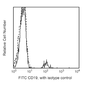
- An isotype control should be used at the same concentration as the antibody of interest.
- Please refer to http://regdocs.bd.com to access safety data sheets (SDS).
- This product is provided under an Agreement between BIOTIUM and BD Biosciences. This product, and only in the amount purchased by buyer, may be used solely for buyer’s own internal research, in a manner consistent with the accompanying product literature. No other right to use, sell or otherwise transfer (a) this product, or (b) its components is hereby granted expressly, by implication or by estoppel. This product is for research use only. Diagnostic uses require a separate license from Biotium, Inc. For information on purchasing a license to this product including for purposes other than research, contact Biotium, Inc., 3159 Corporate Place, Hayward, CA 94545, Tel: (510) 265-1027. Fax: (510) 265-1352. Email: btinfo@biotium.com.
- Alexa Fluor™ is a trademark of Life Technologies Corporation.
Companion Products


The CD1d42 monoclonal antibody recognizes CD1d. Cell surface CD1d is structurally homologous to Class I MHC molecules. It consists of a glycosylated type I transmembrane α chain (43-49 kDa) that is non-covalently associated with β2-microglobulin. CD1d is a member of the CD1 family of molecules, which belong to the immunoglobulin superfamily. Sequence homology data classifies the CD1 molecules into two groups. Group 1 includes CD1a, CD1b and CD1c molecules; group 2 includes CD1d molecules and their homologs in other species. CD1d is expressed on cortical thymocytes, B cells, dendritic cells, monocytes, and some nonlymphoid cells including intestinal epithelial cells, hepatocytes and keratinocytes. It is not expressed on resting mature T cells. Studies suggest that CD1d participates in lipid antigen presentation to CD1d-restricted NKT cells.
The antibody was conjugated to BD Horizon Red 718, which has been developed exclusively for BD Biosciences as a better alternative to Alexa Fluor® 700. BD Horizon Red 718 can be excited by the red laser (628 – 640 nm) and, with an Em Max around 718 nm, it can be detected using a 730/45 nm filter. Due to similar excitation and emission properties, we do not recommend using R718 in combination with APC-R700 or Alexa Fluor® 700.

Development References (6)
-
Exley M, Garcia J, Wilson SB, et al. CD1d structure and regulation on human thymocytes, peripheral blood T cells, B cells and monocytes. Immunology. 2000; 100(1):37-47. (Immunogen: Flow cytometry, Immunohistochemistry, Immunoprecipitation). View Reference
-
Hong S, Scherer DC, Singh N. Lipid antigen presentation in the immune system: lessons learned from CD1d knockout mice. Immunol Rev. 1999; 169:31-44. (Biology). View Reference
-
Kishimoto T. Tadamitsu Kishimoto .. et al., ed. Leucocyte typing VI : white cell differentiation antigens : proceedings of the sixth international workshop and conference held in Kobe, Japan, 10-14 November 1996. New York: Garland Pub.; 1997.
-
Ronger-Savle S, Valladeau J, Claudy A, et al. TGFbeta inhibits CD1d expression on dendritic cells. J Invest Dermatol. 2005; 124(1):116-118. (Clone-specific: Flow cytometry). View Reference
-
Somnay-Wadgaonkar K, Nusrat A, Kim HS. Immunolocalization of CD1d in human intestinal epithelial cells and identification of a beta2-microglobulin-associated form. 1999; 11(3):383-392. (Biology). View Reference
-
Zola H, Swart B, Nicholson I, Voss E. CD1a–e. In: Zola H. Leukocyte and Stromal Cell Molecules : the CD Markers. Hoboken, N.J.: Wiley-Liss; 2007:42-43.
Please refer to Support Documents for Quality Certificates
Global - Refer to manufacturer's instructions for use and related User Manuals and Technical data sheets before using this products as described
Comparisons, where applicable, are made against older BD Technology, manual methods or are general performance claims. Comparisons are not made against non-BD technologies, unless otherwise noted.
For Research Use Only. Not for use in diagnostic or therapeutic procedures.