-
Reagents
- Flow Cytometry Reagents
-
Western Blotting and Molecular Reagents
- Immunoassay Reagents
-
Single-Cell Multiomics Reagents
- BD® OMICS-Guard Sample Preservation Buffer
- BD® AbSeq Assay
- BD® Single-Cell Multiplexing Kit
- BD Rhapsody™ ATAC-Seq Assays
- BD Rhapsody™ Whole Transcriptome Analysis (WTA) Amplification Kit
- BD Rhapsody™ TCR/BCR Next Multiomic Assays
- BD Rhapsody™ Targeted mRNA Kits
- BD Rhapsody™ Accessory Kits
- BD® OMICS-One Protein Panels
-
Functional Assays
-
Microscopy and Imaging Reagents
-
Cell Preparation and Separation Reagents
-
- BD® OMICS-Guard Sample Preservation Buffer
- BD® AbSeq Assay
- BD® Single-Cell Multiplexing Kit
- BD Rhapsody™ ATAC-Seq Assays
- BD Rhapsody™ Whole Transcriptome Analysis (WTA) Amplification Kit
- BD Rhapsody™ TCR/BCR Next Multiomic Assays
- BD Rhapsody™ Targeted mRNA Kits
- BD Rhapsody™ Accessory Kits
- BD® OMICS-One Protein Panels
- Spain (English)
-
Change country/language
Old Browser
This page has been recently translated and is available in French now.
Looks like you're visiting us from United States.
Would you like to stay on the current country site or be switched to your country?
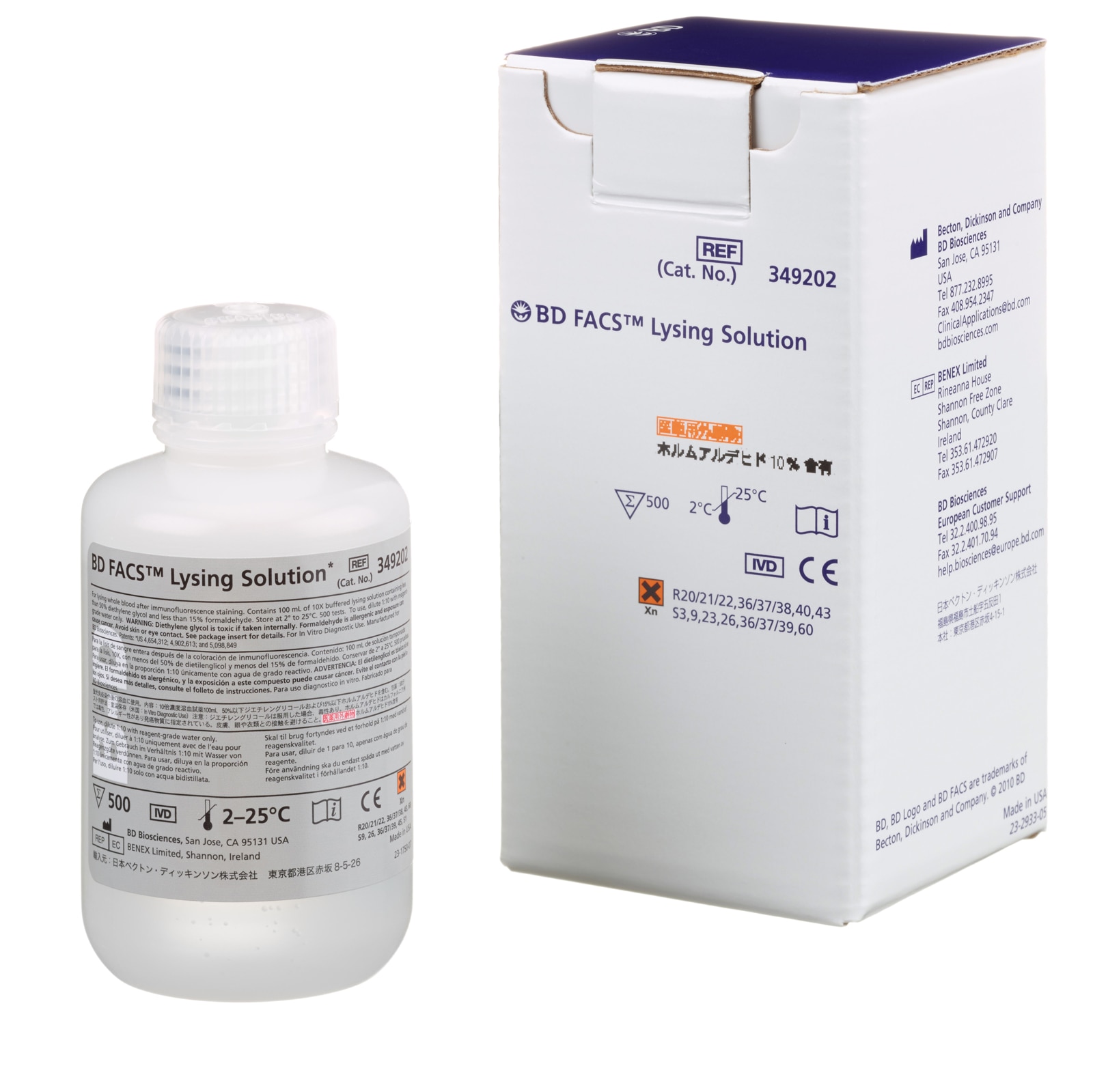
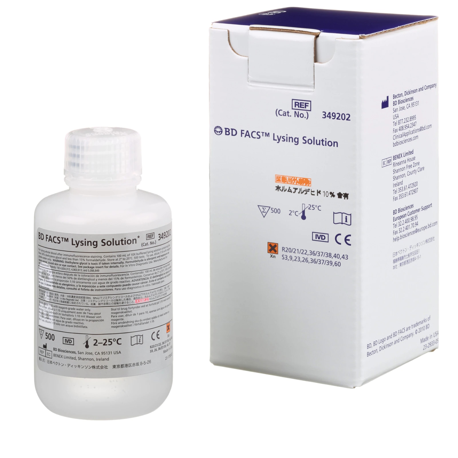
BD FACS™ Lysing Solution 10X Concentrate


Regulatory Status Legend
Any use of products other than the permitted use without the express written authorization of Becton, Dickinson and Company is strictly prohibited.
Product Details
Description
BD FACS™ Lysing solution is intended for lysing red blood cells following direct immunofluorescence staining of human peripheral blood cells with monoclonal antibodies prior to flow cytometric analysis.
BD FACS™ Lysing solution is appropriate for use with reagents such as BD Tritest™ or BD Simultest™ reagents and a suitably equipped flow cytometer. It may be used in both lyse/wash and lyse/no-wash procedures.
Preparation And Storage
BD FACS™ Lysing solution (10X) is stable until the expiration date shown on the bottle label when stored as directed. Do not use this reagent if discoloration occurs or a precipitate forms.
Development References (14)
-
Ashmore LM, Shopp GM, Edwards BS. Lymphocyte subset analysis by flow cytometry. Comparison of three different staining techniques and effects of blood storage. J Immunol Methods. 1989; 118(2):209-215. (Biology). View Reference
-
Centers for Disease Control. Update: universal precautions for prevention of transmission of human immunodeficiency virus, hepatitis B virus, and other bloodborne pathogens in healthcare settings. MMWR. 1988; 37:377-388. (Biology).
-
Clinical Applications of Flow Cytometry: Quality Assurance and Immunophenotyping of Lymphocytes: Approved Guideline. H42-A2. 2007. (Biology).
-
Clinical and Laboratory Standards Institute. 2005. (Biology).
-
De Paoli P, Reitano M, Battistin S, Castiglia C, Santini G. Enumeration of human lymphocyte subsets by monoclonal antibodies and flow cytometry: a comparative study using whole blood or mononuclear cells separated by density gradient centrifugation. J Immunol Methods. 1984; 72(2):349-353. (Biology). View Reference
-
Defining, Establishing, and Verifying Reference Intervals in the Clinical Laboratory; Approved Guideline—Third Edition. C28-A3. 2008. (Biology).
-
Jackson A. Basic phenotyping of lymphocytes: selection and testing of reagents and interpretation of data. Clin Immunol Newslett. 1990; 10:43-55. (Biology).
-
Kidd P, Vogt R. Report of the workshop on the evaluation of T-cell subsets during HIV infection and AIDS. Clin Immunol Immunopathol. 1989; 52:3-9. (Biology).
-
Landay AL, Muirhead KA. Procedural guidelines for performing immunophenotyping by flow cytometry. Clin Immunol Immunopath. 1989; 52:48-60. (Biology).
-
Nicholson JK, Rao PE, Calvelli T, et al. Artifactual staining of monoclonal antibodies in two-color combinations is due to an immunoglobulin in the serum and plasma. Cytometry. 1994; 18:140-146. (Biology).
-
Prince HE, Hirji K, Waldbeser LS, Plaeger-Marshall S, Kleinman S, Lanier LL. Influence of racial background on the distribution of T-cell subsets and Leu 11-positive lymphocytes in healthy blood donors. Diagn Immunol. 1985; 3(1):33-37. (Biology).
-
Procedures for the Collection of Diagnostic Blood Specimens by Venipuncture; Approved Standard—Sixth Edition. H3-A6. 2007. (Biology).
-
Renzi P, Ginns LC. Analysis of T cell subsets in normal adults. Comparison of whole blood lysis technique to Ficoll-Hypaque separation by flow cytometry. J Immunol Methods. 1987; 98(1):53-56. (Biology). View Reference
-
Romeu MA, Mestre M, González L, et al. Lymphocyte immunophenotyping by flow cytometry in normal adults: comparison of fresh whole blood lysis technique, Ficoll-Paque separation and cryopreservation. J Immunol Methods. 1992; 154:44022. (Biology).
Please refer to Support Documents for Quality Certificates
Global - Refer to manufacturer's instructions for use and related User Manuals and Technical data sheets before using this products as described
Comparisons, where applicable, are made against older BD Technology, manual methods or are general performance claims. Comparisons are not made against non-BD technologies, unless otherwise noted.
For In Vitro Diagnostics Use.
Documents are subject to revision without notice. Please verify you have the correct revision of the document, and always refer back to BD's eIFU website for the latest and most up to date information.