
Preparation And Storage
Recommended Assay Procedures
BD® CompBeads can be used as surrogates to assess fluorescence spillover (Compensation). When fluorochrome-conjugated antibodies are bound to BD® CompBeads, they have spectral properties very similar to cells. However, for some fluorochromes there can be small differences in spectral emissions compared to cells, resulting in spillover values that differ when compared to biological controls. It is strongly recommended that when using a reagent for the first time, users compare the spillover on cells and BD® CompBeads. This will ensure that BD® CompBeads are appropriate for your specific cellular application.
For optimal and reproducible results, BD Horizon Brilliant™ Stain Buffer should be used anytime BD Horizon Brilliant™ dyes are used in a multicolor flow cytometry panel. Fluorescent dye interactions may cause staining artifacts which may affect data interpretation. The BD Horizon Brilliant™ Stain Buffer was designed to minimize these interactions. When BD Horizon Brilliant™ Stain Buffer is used in in the multicolor panel, it should also be used in the corresponding compensation controls for all dyes to achieve the most accurate compensation. For the most accurate compensation, compensation controls created with either cells or beads should be exposed to BD Horizon Brilliant™ Stain Buffer for the same length of time as the corresponding multicolor panel. More information can be found in the Technical Data Sheet of the BD Horizon Brilliant™ Stain Buffer (Cat. No. /566349) or the BD Horizon Brilliant™ Stain Buffer Plus (Cat. No. 566385).
Product Notices
- Please refer to www.bdbiosciences.com/us/s/resources for technical protocols.
- Source of all serum proteins is from USDA inspected abattoirs located in the United States.
- Caution: Sodium azide yields highly toxic hydrazoic acid under acidic conditions. Dilute azide compounds in running water before discarding to avoid accumulation of potentially explosive deposits in plumbing.
- This reagent has been pre-diluted for use at the recommended Volume per Test. We typically use 1 × 10^6 cells in a 100-µl experimental sample (a test).
- For fluorochrome spectra and suitable instrument settings, please refer to our Multicolor Flow Cytometry web page at www.bdbiosciences.com/colors.
- An isotype control should be used at the same concentration as the antibody of interest.
- BD Horizon Brilliant Ultraviolet 395 is covered by one or more of the following US patents: 8,158,444; 8,575,303; 8,354,239.
- BD Horizon Brilliant Stain Buffer is covered by one or more of the following US patents: 8,110,673; 8,158,444; 8,575,303; 8,354,239.
- Please refer to http://regdocs.bd.com to access safety data sheets (SDS).
Companion Products


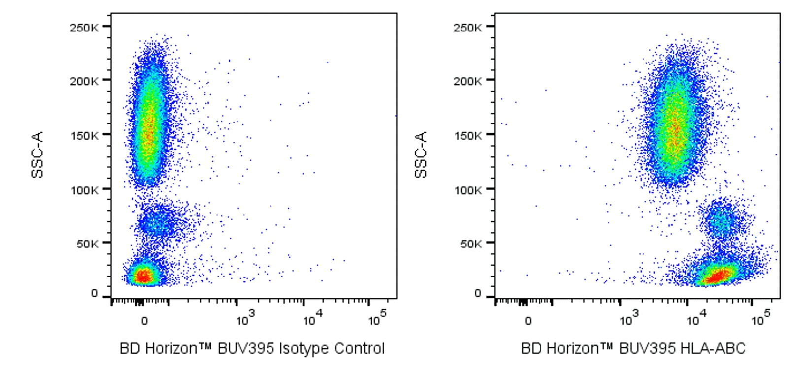
The W6/32 monoclonal antibody recognizes a monomorphic epitope expressed on native β2 microglobulin (β2m)-associated Major Histocompatibility Complex (MHC) class I molecules: Human Leucocyte Antigen (HLA)-A, HLA-B, and HLA-C (HLA-ABC). HLA class I molecules are heterodimers comprised of an ~40-45 kDa, highly polymorphic transmembrane α heavy chain, a type I glycoprotein that is noncovalently-associated with an invariant β2-microglobulin (β2m) light chain. The N-terminal extracellular region of the HLA class I heavy chain is comprised of three domains (α1, α2, and α3). The α1 and α2 domains form a closed antigen-binding groove that accommodates 8-10 aa-peptide antigens. β2m non-covalently associates with the α3 heavy chain domain and promotes HLA class I stability. The W6/32 antibody recognizes a conformational epitope on the HLA class I heavy chain. HLA Class I antigens are normally expressed on most nucleated cells. Their expression is upregulated on activated cells or cells responding to various agents including proinflammatory cytokines or mediators. Reduced HLA Class I expression is found on certain virus-infected or tumor cells. HLA class I antigens expressed on thymic epithelial cells are involved in the positive and negative selection of CD8+ T cell precursors which determines their TCR repertoire during T cell maturation. In the periphery, these HLA Class I antigens serve to either present endogenous antigens or cross-present exogenous antigens for the generation of effector and memory CD8+ T cell responses. Target cell HLA Class I antigens can serve as ligands for inhibitory receptors expressed on NK cells and CD8+ T cells and suppress their cytotoxic responses. Human HLA class I molecules play critical roles in cell-mediated immune responses and tumor surveillance as well as tolerance to self-antigens.
The antibody was conjugated to BD Horizon BUV395 which has been exclusively developed by BD Biosciences as an optimal dye for use on a 355 nm laser equipped instrument. With an Ex Max at 348 nm and an Em Max at 395 nm, this dye has virtually no spillover into any other detector. BD Horizon BUV395 can be excited with a 355 nm laser and detected with a 379/28 filter.

Development References (5)
-
Barnstable CJ, Bodmer WF, Brown G, et al. Production of monoclonal antibodies to group A erythrocytes, HLA and other human cell surface antigens-new tools for genetic analysis.. Cell. 1978; 14(1):9-20. (Immunogen: Cytotoxicity, Immunoprecipitation, Radioimmunoassay). View Reference
-
Margulies DH, Natarajan K, Rossjohn J, McCluskey J. Major Histocompatibility Complex and Its Proteins. In: Paul WE. Paul WE, ed. Fundamental Immunology 7th Edition. Philadelphia: Lippincott Williams & Wilkins; 2013:487-523.
-
Parham P, Barnstable CJ, Bodmer WF. Use of a monoclonal antibody (W6/32) in structural studies of HLA-A,B,C, antigens.. J Immunol. 1979; 123(1):342-9. (Clone-specific: Immunoaffinity chromatography, Immunoprecipitation, Radioimmunoassay). View Reference
-
Shields MJ, Ribaudo RK. Mapping of the monoclonal antibody W6/32: sensitivity to the amino terminus of beta2-microglobulin.. Tissue Antigens. 1998; 51(5):567-70. (Clone-specific: Flow cytometry). View Reference
-
Wooden SL, Kalb SR, Cotter RJ, Soloski MJ. Cutting edge: HLA-E binds a peptide derived from the ATP-binding cassette transporter multidrug resistance-associated protein 7 and inhibits NK cell-mediated lysis.. J Immunol. 2005; 175(3):1383-7. (Clone-specific: Flow cytometry, Functional assay, Inhibition). View Reference
Please refer to Support Documents for Quality Certificates
Global - Refer to manufacturer's instructions for use and related User Manuals and Technical data sheets before using this products as described
Comparisons, where applicable, are made against older BD Technology, manual methods or are general performance claims. Comparisons are not made against non-BD technologies, unless otherwise noted.
For Research Use Only. Not for use in diagnostic or therapeutic procedures.