-
Reagents
- Flow Cytometry Reagents
-
Western Blotting and Molecular Reagents
- Immunoassay Reagents
-
Single-Cell Multiomics Reagents
- BD® OMICS-Guard Sample Preservation Buffer
- BD® AbSeq Assay
- BD® Single-Cell Multiplexing Kit
- BD Rhapsody™ ATAC-Seq Assays
- BD Rhapsody™ Whole Transcriptome Analysis (WTA) Amplification Kit
- BD Rhapsody™ TCR/BCR Next Multiomic Assays
- BD Rhapsody™ Targeted mRNA Kits
- BD Rhapsody™ Accessory Kits
- BD® OMICS-One Protein Panels
- BD OMICS-One™ WTA Next Assay
-
Functional Assays
-
Microscopy and Imaging Reagents
-
Cell Preparation and Separation Reagents
Old Browser
This page has been recently translated and is available in French now.
Looks like you're visiting us from {countryName}.
Would you like to stay on the current location site or be switched to your location?
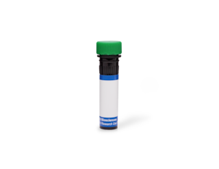
BD Transduction Laboratories™ Purified Mouse Anti-Rat TGN38
Clone 2/TGN38 (RUO)


Western blot analysis of TGN38 on a rat cerebum lysate. Lane 1: 1:250, lane 2: 1: 500, lane 3: 1: 1000 dilution of the mouse anti- rat TGN38 antibody.

Immunofluorescence staining of L6 cells (Rat skeletal muscle myoblasts; ATCC CRL-1458).


Regulatory Status Legend
Any use of products other than the permitted use without the express written authorization of Becton, Dickinson and Company is strictly prohibited.
Preparation And Storage
Recommended Assay Procedures
Western blot: Please refer to http://www.bdbiosciences.com/pharmingen/protocols/Western_Blotting.shtml
Product Notices
- Since applications vary, each investigator should titrate the reagent to obtain optimal results.
- Please refer to www.bdbiosciences.com/us/s/resources for technical protocols.
- Source of all serum proteins is from USDA inspected abattoirs located in the United States.
- Caution: Sodium azide yields highly toxic hydrazoic acid under acidic conditions. Dilute azide compounds in running water before discarding to avoid accumulation of potentially explosive deposits in plumbing.
Companion Products


Newly synthesized proteins exit the ER and move to the cis-Golgi network (CGN) where they traverse the cis-medial and trans-cisternae before reaching the trans-Golgi network (TGN). N-linked oligosaccharide processing occurs in the TGN, and proteins are sorted to the plasma membrane, lysosomes, endosomes, and secretory granules. TGN38 is a type I integral membrane protein primarily localized to the TGN. It is involved in the sorting of nascent proteins into individual carrier vesicles for transport to appropriate destinations. It is thought to heterodimerize with TGN41 and participate in exocytic budding from the TGN. TGN38 has a molecular weight of 85 to 95 kDa. The core polypeptide represents approximately 38 kDa, while the remainder is accounted for by N- and O-linked oligosaccharide chains. A 286 aa N-terminal lumenal domain, a 21 aa membrane spanning domain, and a 33 aa C-terminal cytoplasmic tail comprise the structure of TGN38. The cytoplasmic tail contains a tyrosine-based motif, YQRL, that is thought to be involved in TGN localization. Therefore, TGN38 mediates the localization of various proteins to the TGN and serves as a TGN retrieval signal.
This antibody is routinely tested by western blot analysis. Other applications were tested at BD Biosciences Pharmingen during antibody development only or reported in the literature.
Development References (5)
-
Humphrey JS, Peters PJ, Yuan LC, Bonifacino JS. Localization of TGN38 to the trans-Golgi network: involvement of a cytoplasmic tyrosine-containing sequence. J Cell Biol. 1993; 120(5):1123-1135. (Biology). View Reference
-
Luzio JP, Brake B, Banting G, Howell KE, Braghetta P, Stanley KK. Identification, sequencing and expression of an integral membrane protein of the trans-Golgi network (TGN38). Biochem J. 1990; 270(1):97-102. (Biology). View Reference
-
Mary S, Charrasse S, Meriane M, et al. Biogenesis of N-cadherin-dependent cell-cell contacts in living fibroblasts is a microtubule-dependent kinesin-driven mechanism. Mol Biol Cell. 2002; 13(1):285-301. (Biology: Immunofluorescence). View Reference
-
Ozawa K, Kondo T, Hori O. Expression of the oxygen-regulated protein ORP150 accelerates wound healing by modulating intracellular VEGF transport. J Clin Invest. 2001; 108(1):39-40. (Biology: Western blot). View Reference
-
Xu L, Shen Y, Joseph T, et al. Mitogenic phospholipase D activity is restricted to caveolin-enriched membrane microdomains. Biochem Biophys Res Commun. 2000; 273(1):77-83. (Biology: Western blot). View Reference
Please refer to Support Documents for Quality Certificates
Global - Refer to manufacturer's instructions for use and related User Manuals and Technical data sheets before using this products as described
Comparisons, where applicable, are made against older BD Technology, manual methods or are general performance claims. Comparisons are not made against non-BD technologies, unless otherwise noted.
For Research Use Only. Not for use in diagnostic or therapeutic procedures.