





.png)


Preparation And Storage


Product Notices
- Since applications vary, each investigator should titrate the reagent to obtain optimal results.
- An isotype control should be used at the same concentration as the antibody of interest.
- Caution: Sodium azide yields highly toxic hydrazoic acid under acidic conditions. Dilute azide compounds in running water before discarding to avoid accumulation of potentially explosive deposits in plumbing.
- Although hamster immunoglobulin isotypes have not been well defined, BD Biosciences Pharmingen has grouped Armenian and Syrian hamster IgG monoclonal antibodies according to their reactivity with a panel of mouse anti-hamster IgG mAbs. A table of the hamster IgG groups, Reactivity of Mouse Anti-Hamster Ig mAbs, may be viewed at http://www.bdbiosciences.com/documents/hamster_chart_11x17.pdf.
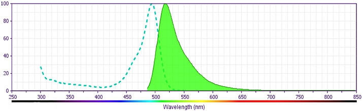
- For fluorochrome spectra and suitable instrument settings, please refer to our Multicolor Flow Cytometry web page at www.bdbiosciences.com/colors.
- Please refer to www.bdbiosciences.com/us/s/resources for technical protocols.
Companion Products


.png?imwidth=320)
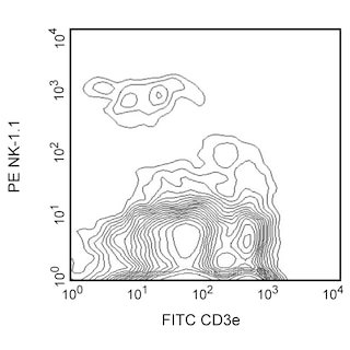
The HL3 monoclonal antibody specifically binds to the integrin αx chain of gp150, 95 (CD11c/CD18). CD11c is expressed on dendritic cells, CD4- CD8+ intestinal intraepithelial lymphocytes (IEL) and some NK cells. It is upregulated on IEL and lymph-node T cells following in vivo activation. Cells of the monocyte/macrophage lineage have been reported to express low levels of CD11c. CD11c plays a role in binding of iC3b.
Development References (8)
-
Barclay NA, Brown MH, Birkeland ML, et al, ed. The Leukocyte Antigen FactsBook. San Diego, CA: Academic Press; 1997.
-
Burt BM, Plitas G, Stableford JA, et al. CD11c identifies a subset of murine liver natural killer cells that responds to adenoviral hepatitis. J Leukoc Biol. 2008; 84(4):1039-1046. (Clone-specific). View Reference
-
Fagarasan S, Muramatsu M, Suzuki K, Nagaoka H, Hiai H, Honjo T. Critical roles of activation-induced cytidine deaminase in the homeostasis of gut flora. Science. 2002; 298(5597):1424-1427. (Clone-specific: Immunofluorescence). View Reference
-
Gao JX, Liu X, Wen J, et al. Differentiation of monocytic cell clones into CD8 alpha+ dendritic cells (DC) suggests that monocytes can be direct precursors for both CD8 alpha+ and CD8 alpha- DC in the mouse. J Immunol. 2003; 170(12):5927-5935. (Biology). View Reference
-
Huleatt JW, Lefrançois L. Antigen-driven induction of CD11c on intestinal intraepithelial lymphocytes and CD8+ T cells in vivo.. J Immunol. 1995; 154(11):5684-93. (Immunogen: Immunoprecipitation). View Reference
-
Larson RS, Springer TA. Structure and function of leukocyte integrins. Immunol Rev. 1990; 114:181-217. (Biology). View Reference
-
Maraskovsky E, Brasel K, Teepe M, et al. Dramatic increase in the numbers of functionally mature dendritic cells in Flt3 ligand-treated mice: multiple dendritic cell subpopulations identified. J Exp Med. 1996; 184(5):1953-1962. (Biology). View Reference
-
Pulendran B, Lingappa J, Kennedy MK, et al. Developmental pathways of dendritic cells in vivo: distinct function, phenotype, and localization of dendritic cell subsets in FLT3 ligand-treated mice. J Immunol. 1997; 159(5):2222-2231. (Clone-specific: Immunohistochemistry). View Reference
Please refer to Support Documents for Quality Certificates
Global - Refer to manufacturer's instructions for use and related User Manuals and Technical data sheets before using this products as described
Comparisons, where applicable, are made against older BD Technology, manual methods or are general performance claims. Comparisons are not made against non-BD technologies, unless otherwise noted.
For Research Use Only. Not for use in diagnostic or therapeutic procedures.
