





BD Horizon™ BV605 Mouse Anti-Human TSLP Receptor
Clone 1F11/TSLPR (also known as 1F11 and AB81_85.1F11)
(RUO)




Preparation And Storage


Product Notices
- This reagent has been pre-diluted for use at the recommended Volume per Test. We typically use 1 × 10^6 cells in a 100-µl experimental sample (a test).
- An isotype control should be used at the same concentration as the antibody of interest.
- Please refer to www.bdbiosciences.com/us/s/resources for technical protocols.
- Please observe the following precautions: Absorption of visible light can significantly alter the energy transfer occurring in any tandem fluorochrome conjugate; therefore, we recommend that special precautions be taken (such as wrapping vials, tubes, or racks in aluminum foil) to prevent exposure of conjugated reagents, including cells stained with those reagents, to room illumination.
- Caution: Sodium azide yields highly toxic hydrazoic acid under acidic conditions. Dilute azide compounds in running water before discarding to avoid accumulation of potentially explosive deposits in plumbing.
- For fluorochrome spectra and suitable instrument settings, please refer to our Multicolor Flow Cytometry web page at www.bdbiosciences.com/colors.
- Although every effort is made to minimize the lot-to-lot variation in the efficiency of the fluorochrome energy transfer, differences in the residual emission from BD Horizon™ BV421 may be observed. Therefore, we recommend that individual compensation controls be performed for every BD Horizon™ BV605 conjugate.
- CF™ is a trademark of Biotium, Inc.
Companion Products


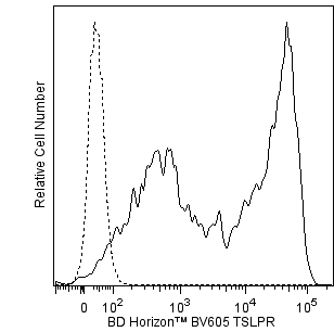
The 1F11/TSLPR monoclonal antibody specifically binds to Thymic Stromal Lymphopoietin Receptor (TSLPR). TSLPR is a member of the hematopoietin receptor superfamily and is also known as Cytokine Receptor-like Factor 2 (CRL2, CRLF2Y, CRLF2). The functional TSLPR complex consists of two subunits, TSLPR and the alpha subunit of the Interleukin-7 Receptor (IL-7Rα). Analysis of the TSLPR reveals sequence similarity with the common cytokine receptor gamma chain (γc; CD132). Functional TSLPRs are expressed by epithelial cells and a variety of hematopoietic cell types, including thymocytes, T cells, B cells, natural killer T cells, monocytes, macrophages, basophils, and dendritic cells (DC). Recent studies indicate that TSLP can activate multiple STAT (Signal Transducer and Activator of Transcription) signaling proteins. TSLP enhances the maturation and viability of DC. It strongly induces DC expression of the CD40 and CD80 costimulatory molecules and chemokines, e.g., TARC (Thymus and activation-regulated chemokine; CCL17) that can attract Th2 effector cells. TSLP supports B cell development. TSLP costimulates the proliferation of naïve T cells in the presence of mature DC. TSLP is also able to increase the sensitivity of T cell receptor-activated CD4+ T cells to low doses of IL-2. In the presence of TSLP, the acute myeloid leukemia-derived cell line, MUTZ-3, shows induced growth and reduced apoptosis. CRLF2 deregulated gene expression is thought to be involved in lymphoid transformation in B-cell precursor acute lymphoblastic leukemia. The 1F11/TSLPR antibody is reportedly a neutralizing antibody.
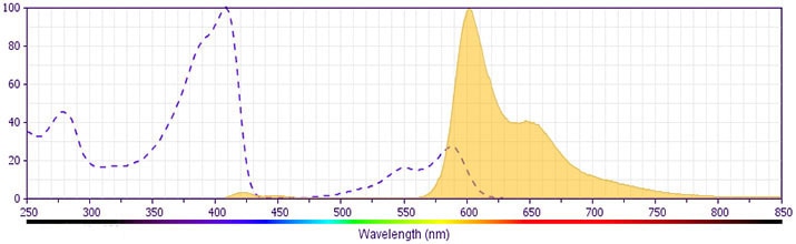
This antibody is conjugated to BD Horizon BV605 which is part of the BD Horizon Brilliant™ Violet family of dyes. With an Ex Max of 407-nm and Em Max of 602-nm, BD Horizon BV605 can be excited by a violet laser and detected with a standard 610/20-nm filter set. BD Horizon BV605 is a tandem fluorochrome of BD Horizon BV421 and an acceptor dye with an Em max at 605-nm. Due to the excitation of the acceptor dye by the green (532 nm) and yellow-green (561 nm) lasers, there will be significant spillover into the PE and BD Horizon PE-CF594 detectors off the green or yellow-green lasers. BD Horizon BV605 conjugates are very bright, often exhibiting brightness equivalent to PE conjugates and can be used as a third color off of the violet laser.
For optimal and reproducible results, BD Horizon Brilliant Stain Buffer should be used anytime two or more BD Horizon Brilliant dyes are used in the same experiment. Fluorescent dye interactions may cause staining artifacts which may affect data interpretation. The BD Horizon Brilliant Stain Buffer was designed to minimize these interactions. More information can be found in the Technical Data Sheet of the BD Horizon Brilliant Stain Buffer (Cat. No. 563794).
Development References (10)
-
Arima K, Watanabe N, Hanabuchi S, Chang M, Sun SC, Liu YJ. Distinct signal codes generate dendritic cell functional plasticity. Sci Signal. 2010; 3(105):ra4. (Clone-specific: Neutralization). View Reference
-
Ito T, Liu YJ, Arima K. Cellular and molecular mechanisms of TSLP function in human allergic disorders--TSLP programs the "Th2 code" in dendritic cells. Allergol Int. 2012; 61(1):35-43. (Biology). View Reference
-
Lu N, Wang YH, Arima K, Hanabuchi S, Liu YJ. TSLP and IL-7 use two different mechanisms to regulate human CD4+ T cell homeostasis. J Exp Med. 2009; 206(10):2111-2119. (Biology). View Reference
-
Pedroza-Gonzalez A, Xu K, Wu TC, et al. Thymic stromal lymphopoietin fosters human breast tumor growth by promoting type 2 inflammation. J Exp Med. 2011; 208(3):479-490. (Clone-specific: Neutralization). View Reference
-
Quentmeier H, Drexler HG, Fleckenstein D, et al. Cloning of human thymic stromal lymphopoietin (TSLP) and signaling mechanisms leading to proliferation. Leukemia. 2001; 15(8):1286-1292. (Biology). View Reference
-
Reche PA, Soumelis V, Gorman DM, et al. Human thymic stromal lymphopoietin preferentially stimulates myeloid cells. J Immunol. 2001; 167(1):336-343. (Biology). View Reference
-
Rochman I, Watanabe N, Arima K, Liu YJ, Leonard WJ. Cutting edge: direct action of thymic stromal lymphopoietin on activated human CD4+ T cells. J Immunol. 2007; 178(11):6720-6724. (Biology). View Reference
-
Russell LJ, Capasso M, Vater I, et al. Deregulated expression of cytokine receptor gene, CRLF2, is involved in lymphoid transformation in B-cell precursor acute lymphoblastic leukemia. Blood. 2009; 114(13):2688-2698. (Biology). View Reference
-
Tedla N, Bandeira-Melo C, Tassinari P, et al. Activation of human eosinophils through leukocyte immunoglobulin-like receptor 7. Proc Natl Acad Sci U S A. 2003; 100(3):1174-1179. (Biology). View Reference
-
Ziegler SF. Thymic stromal lymphopoietin and allergic disease. J Allergy Clin Immunol. 2012; 130(4):845-852. (Biology). View Reference
Please refer to Support Documents for Quality Certificates
Global - Refer to manufacturer's instructions for use and related User Manuals and Technical data sheets before using this products as described
Comparisons, where applicable, are made against older BD Technology, manual methods or are general performance claims. Comparisons are not made against non-BD technologies, unless otherwise noted.
For Research Use Only. Not for use in diagnostic or therapeutic procedures.
