







Preparation And Storage


Recommended Assay Procedures
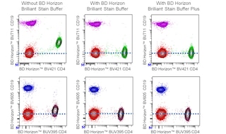
For optimal and reproducible results, BD Horizon Brilliant Stain Buffer should be used anytime two or more BD Horizon Brilliant dyes are used in the same experiment. Fluorescent dye interactions may cause staining artifacts which may affect data interpretation. The BD Horizon Brilliant Stain Buffer was designed to minimize these interactions. More information can be found in the Technical Data Sheet of the BD Horizon Brilliant Stain Buffer (Cat. No. 563794/566349) or the BD Horizon Brilliant Stain Buffer Plus (Cat. No. 566385).
Product Notices
- This reagent has been pre-diluted for use at the recommended Volume per Test. We typically use 1 × 10^6 cells in a 100-µl experimental sample (a test).
- Source of all serum proteins is from USDA inspected abattoirs located in the United States.
- An isotype control should be used at the same concentration as the antibody of interest.
- Caution: Sodium azide yields highly toxic hydrazoic acid under acidic conditions. Dilute azide compounds in running water before discarding to avoid accumulation of potentially explosive deposits in plumbing.
- For fluorochrome spectra and suitable instrument settings, please refer to our Multicolor Flow Cytometry web page at www.bdbiosciences.com/colors.
- BD Horizon Brilliant Violet 510 is covered by one or more of the following US patents: 8,575,303; 8,354,239.
- BD Horizon Brilliant Stain Buffer is covered by one or more of the following US patents: 8,110,673; 8,158,444; 8,575,303; 8,354,239.
- Species cross-reactivity detected in product development may not have been confirmed on every format and/or application.
- Please refer to www.bdbiosciences.com/us/s/resources for technical protocols.
Companion Products




The G46-6 monoclonal antibody specifically binds to HLA-DR, a major histocompatibility complex (MHC) class II antigen. HLA-DR antigens are encoded by genes within the Human Leukocyte Antigen (HLA) Complex located on chromosome 6. HLA-DR is a transmembrane heterodimeric glycoprotein composed of an α chain (36 kDa) and a β subunit (27 kDa) expressed primarily on antigen presenting cells: B cells, dendritic cells, monocytes, macrophages, and thymic epithelial cells. HLA-DR is also expressed on activated T cells. This molecule plays a major role in mediating cellular interactions during antigen presentation to CD4-positive T cells.
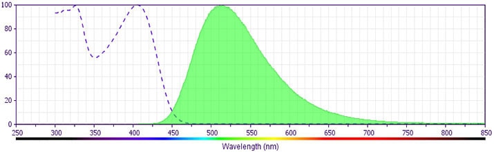
The antibody was conjugated to BD Horizon™ BV510 which is part of the BD Horizon Brilliant™ Violet family of dyes. With an Ex Max of 405-nm and Em Max at 510-nm, BD Horizon™ BV510 can be excited by the violet laser and detected in the BD Horizon™ V500 (525/50-nm) filter set. BD Horizon™ BV510 conjugates are useful for the detection of dim markers off the violet laser.
Development References (5)
-
Dieckmann D, Plottner H, Berchtold S, Berger T, Schuler G. Ex vivo isolation and characterization of CD4(+)CD25(+) T cells with regulatory properties from human blood. J Exp Med. 2001; 193(11):1303-1310. (Clone-specific: Flow cytometry). View Reference
-
Ibisch C, Pradal G, Bach JM, Lieubeau B. Functional canine dendritic cells can be generated in vitro from peripheral blood mononuclear cells and contain a cytoplasmic ultrastructural marker.. J Immunol Methods. 2005; 298(1-2):175-82. (Clone-specific). View Reference
-
Kitani A, Chua K, Nakamura K, Strober W. Activated self-MHC-reactive T cells have the cytokine phenotype of Th3/T regulatory cell 1 T cells. J Immunol. 2000; 165(2):691-702. (Clone-specific: Flow cytometry). View Reference
-
Moran TP, Collier M, McKinnon KP, Davis NL, Johnston RE, Serody JS. A novel viral system for generating antigen-specific T cells. J Immunol. 2008; 175(5):3431-3438. (Clone-specific: Flow cytometry). View Reference
-
Sorg RV, Kogler G, Wernet P. Identification of cord blood dendritic cells as an immature CD11c- population. Blood. 1999; 93(7):2302-2307. (Clone-specific: Flow cytometry). View Reference
Please refer to Support Documents for Quality Certificates
Global - Refer to manufacturer's instructions for use and related User Manuals and Technical data sheets before using this products as described
Comparisons, where applicable, are made against older BD Technology, manual methods or are general performance claims. Comparisons are not made against non-BD technologies, unless otherwise noted.
For Research Use Only. Not for use in diagnostic or therapeutic procedures.
