







Preparation And Storage


Recommended Assay Procedures
For optimal and reproducible results, BD Horizon Brilliant Stain Buffer should be used anytime two or more BD Horizon Brilliant dyes are used in the same experiment. Fluorescent dye interactions may cause staining artifacts which may affect data interpretation. The BD Horizon Brilliant Stain Buffer was designed to minimize these interactions. More information can be found in the Technical Data Sheet of the BD Horizon Brilliant Stain Buffer (Cat. No. 563794).
Product Notices
- This reagent has been pre-diluted for use at the recommended Volume per Test. We typically use 1 × 10^6 cells in a 100-µl experimental sample (a test).
- An isotype control should be used at the same concentration as the antibody of interest.
- Source of all serum proteins is from USDA inspected abattoirs located in the United States.
- Caution: Sodium azide yields highly toxic hydrazoic acid under acidic conditions. Dilute azide compounds in running water before discarding to avoid accumulation of potentially explosive deposits in plumbing.
- BD Horizon Brilliant Violet 480 is covered by one or more of the following US patents: 8,575,303; 8,354,239.
- For fluorochrome spectra and suitable instrument settings, please refer to our Multicolor Flow Cytometry web page at www.bdbiosciences.com/colors.
- Please refer to www.bdbiosciences.com/us/s/resources for technical protocols.
Companion Products



The MI15 monoclonal antibody specifically binds to CD138 (Syndecan-1), an 85-92 kDa single chain transmembrane protein, which is strongly expressed on multiple-myeloma-derived cell lines and malignant plasma cell populations. It is also expressed on pre-B cells, immature B cells, and plasma cells, but not on mature circulating B-lymphocytes. Syndecan-1 is a member of the transmembrane heparan sulfate proteoglycans family. It is also expressed on some non-hematopoietic cells, including embryonic mesenchymal cells, vascular smooth muscle cells, endothelial and neural cells. CD138 binds to many extracellular matrix proteins through its heparan sulfate side-chains, like fibronectin, collagen types I, III, and V, tenascin, thrombospondin, and antithrombin III. It is considered an extracellular matrix receptor that may serve as a co-receptor for fibroblast growth factor and related molecules. Monoclonal antibody MI15 blocks the binding of clone B-B4 but not clone DL-101 (other anti-syndecan-1 antibodies) by flow cytometric analysis.
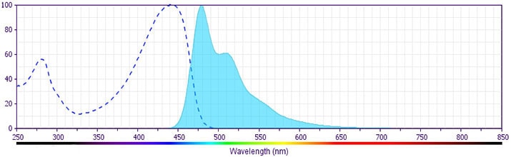
The antibody was conjugated to BD Horizon BV480 which is part of the BD Horizon Brilliant™ Violet family of dyes. With an Ex Max of 436-nm and Em Max at 478-nm, BD Horizon BV480 can be excited by the violet laser and detected in the BD Horizon BV510 (525/40-nm) filter set. BV480 has less spillover into the BV605 detector and, in general, is brighter than BV510.
Development References (6)
-
Barclay NA, Brown MH, Birkeland ML, et al, ed. The Leukocyte Antigen FactsBook. San Diego, CA: Academic Press; 1997.
-
Costes V, Magen V, Legouffe E, et al. The Mi15 monoclonal antibody (anti-syndecan-1) is a reliable marker for quantifying plasma cells in paraffin-embedded bone marrow biopsy specimens. Hum Pathol. 1999; 30(12):1405-1411. (Immunogen: Immunohistochemistry). View Reference
-
Gattei V, Godeas C, Degan M, Rossi FM, Aldinucci D, Pinto A. Characterization of anti-CD138 monoclonal antibodies as tools for investigating the molecular polymorphism of syndecan-1 in human lymphoma cells. Br J Haematol. 1999; 104(1):152-162. (Clone-specific: Flow cytometry, Immunoprecipitation). View Reference
-
Horvathova M, Gaillard JP, Liautard J, et al. Identification of novel and specific antigens of human plasma cells by mAb. In: Schlossman SF. Stuart F. Schlossman .. et al., ed. Leucocyte typing V : white cell differentiation antigens : proceedings of the fifth international workshop and conference held in Boston, USA, 3-7 November, 1993. Oxford: Oxford University Press; 1995:713-714.
-
Wijdenes J, Clément C, Klein B, Dore J-M. CD138 (syndecan-1) Workshop Panel report. In: Kishimoto T. Tadamitsu Kishimoto .. et al., ed. Leucocyte typing VI : white cell differentiation antigens : proceedings of the sixth international workshop and conference held in Kobe, Japan, 10-14 November 1996. New York: Garland Pub.; 1997:249-252.
-
Zola H. Leukocyte and stromal cell molecules : the CD markers. Hoboken, N.J.: Wiley-Liss; 2007.
Please refer to Support Documents for Quality Certificates
Global - Refer to manufacturer's instructions for use and related User Manuals and Technical data sheets before using this products as described
Comparisons, where applicable, are made against older BD Technology, manual methods or are general performance claims. Comparisons are not made against non-BD technologies, unless otherwise noted.
For Research Use Only. Not for use in diagnostic or therapeutic procedures.
