

Preparation And Storage
Recommended Assay Procedures
BD® CompBeads can be used as surrogates to assess fluorescence spillover (compensation). When fluorochrome conjugated antibodies are bound to BD® CompBeads, they have spectral properties very similar to cells. However, for some fluorochromes there can be small differences in spectral emissions compared to cells, resulting in spillover values that differ when compared to biological controls. It is strongly recommended that when using a reagent for the first time, users compare the spillover on cells and BD® CompBeads to ensure that BD® CompBeads are appropriate for your specific cellular application.
For optimal and reproducible results, BD Horizon Brilliant Stain Buffer should be used anytime BD Horizon Brilliant dyes are used in a multicolor flow cytometry panel. Fluorescent dye interactions may cause staining artifacts which may affect data interpretation. The BD Horizon Brilliant Stain Buffer was designed to minimize these interactions. When BD Horizon Brilliant Stain Buffer is used in in the multicolor panel, it should also be used in the corresponding compensation controls for all dyes to achieve the most accurate compensation. For the most accurate compensation, compensation controls created with either cells or beads should be exposed to BD Horizon Brilliant Stain Buffer for the same length of time as the corresponding multicolor panel. More information can be found in the Technical Data Sheet of the BD Horizon Brilliant Stain Buffer (Cat. No. 563794/566349) or the BD Horizon Brilliant Stain Buffer Plus (Cat. No. 566385).
Product Notices
- Please refer to www.bdbiosciences.com/us/s/resources for technical protocols.
- This reagent has been pre-diluted for use at the recommended Volume per Test. We typically use 1 × 10^6 cells in a 100-µl experimental sample (a test).
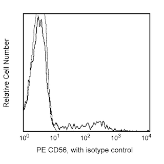
- An isotype control should be used at the same concentration as the antibody of interest.
- Caution: Sodium azide yields highly toxic hydrazoic acid under acidic conditions. Dilute azide compounds in running water before discarding to avoid accumulation of potentially explosive deposits in plumbing.
- For fluorochrome spectra and suitable instrument settings, please refer to our Multicolor Flow Cytometry web page at www.bdbiosciences.com/colors.
- Human donor specific background has been observed in relation to the presence of anti-polyethylene glycol (PEG) antibodies, developed as a result of certain vaccines containing PEG, including some COVID-19 vaccines. We recommend use of BD Horizon Brilliant™ Stain Buffer in your experiments to help mitigate potential background. For more information visit https://www.bdbiosciences.com/en-us/support/product-notices.
- Please refer to http://regdocs.bd.com to access safety data sheets (SDS).
- For U.S. patents that may apply, see bd.com/patents.
Companion Products


The TgMab-2 monoclonal antibody specifically recognizes TIGIT (T cell Immunoreceptor with Ig and ITIM domains) which is also known as Vstm3 (V-set and transmembrane domain-containing 3), Vsig9 (V-set and Ig domain-containing 9) and WUCAM (Washington University Cell Adhesion Molecule). TIGIT is a 30-34 kDa single pass type I transmembrane glycoprotein that belongs to the CD28 family within the Ig superfamily. TIGIT has an extracellular region with a V-type Ig-like domain, transmembrane sequence, and a cytoplasmic domain with an immunoreceptor tyrosine-based inhibitory motif (ITIM). TIGIT is expressed on NK cells and subsets of activated and memory T cells, regulatory T cells (Treg), and T follicular helper (Tfh) cells. TIGIT binds to CD112 (PVRL2/Nectin-2) and CD155 (PVR/Necl-5) that are expressed on dendritic cells (DC), endothelial cells, fibroblasts, and some tumor cells and can induce IL-10 release and inhibition of IL-12 production. Ligand-bound TIGIT downregulates TCR-mediated T cell activation and proliferation and can block NK cell-mediated cytotoxicity.

Development References (3)
-
Downs-Canner S, Berkey S, Delgoffe GM, et al. Suppressive IL-17A(+)Foxp3(+) and ex-Th17 IL-17A(neg)Foxp3(+) Treg cells are a source of tumour-associated Treg cells.. Nat Commun. 2017; 8:14649. (Biology). View Reference
-
Yasuma K, Yasunaga J, Takemoto K, et al. HTLV-1 bZIP Factor Impairs Anti-viral Immunity by Inducing Co-inhibitory Molecule, T Cell Immunoglobulin and ITIM Domain (TIGIT).. PLoS Pathog. 2016; 12(1):e1005372. (Biology). View Reference
-
Yu X, Harden K, Gonzalez LC, Francesco M, et al. The surface protein TIGIT suppresses T cell activation by promoting the generation of mature immunoregulatory dendritic cells. Nat Immunol. 2009; 10(1):48-57. (Biology). View Reference
Please refer to Support Documents for Quality Certificates
Global - Refer to manufacturer's instructions for use and related User Manuals and Technical data sheets before using this products as described
Comparisons, where applicable, are made against older BD Technology, manual methods or are general performance claims. Comparisons are not made against non-BD technologies, unless otherwise noted.
For Research Use Only. Not for use in diagnostic or therapeutic procedures.