

Preparation And Storage
Recommended Assay Procedures
BD® CompBeads can be used as surrogates to assess fluorescence spillover (Compensation). When fluorochrome-conjugated antibodies are bound to BD® CompBeads, they have spectral properties very similar to cells. However, for some fluorochromes there can be small differences in spectral emissions compared to cells, resulting in spillover values that differ when compared to biological controls. It is strongly recommended that when using a reagent for the first time, users compare the spillover on cells and BD® CompBeads. This will ensure that BD® CompBeads are appropriate for your specific cellular application.
For optimal and reproducible results, BD Horizon Brilliant™ Stain Buffer should be used anytime BD Horizon Brilliant™ dyes are used in a multicolor flow cytometry panel. Fluorescent dye interactions may cause staining artifacts which may affect data interpretation. The BD Horizon Brilliant™ Stain Buffer was designed to minimize these interactions. When BD Horizon Brilliant™ Stain Buffer is used in in the multicolor panel, it should also be used in the corresponding compensation controls for all dyes to achieve the most accurate compensation. For the most accurate compensation, compensation controls created with either cells or beads should be exposed to BD Horizon Brilliant™ Stain Buffer for the same length of time as the corresponding multicolor panel. More information can be found in the Technical Data Sheet of the BD Horizon Brilliant™ Stain Buffer (Cat. No. 563794/566349) or the BD Horizon Brilliant™ Stain Buffer Plus (Cat. No. 566385).
Note: When using high concentrations of antibody, background binding of this dye to erythroid cell subsets (mature erythrocytes and precursors) has been observed. For researchers studying these cell populations, or in cases where light scatter gating does not adequately exclude these cells from the analysis, this background may be an important factor to consider when selecting reagents for panel(s).
Product Notices
- Please refer to www.bdbiosciences.com/us/s/resources for technical protocols.
- Caution: Sodium azide yields highly toxic hydrazoic acid under acidic conditions. Dilute azide compounds in running water before discarding to avoid accumulation of potentially explosive deposits in plumbing.
- This reagent has been pre-diluted for use at the recommended Volume per Test. We typically use 1 × 10^6 cells in a 100-µl experimental sample (a test).
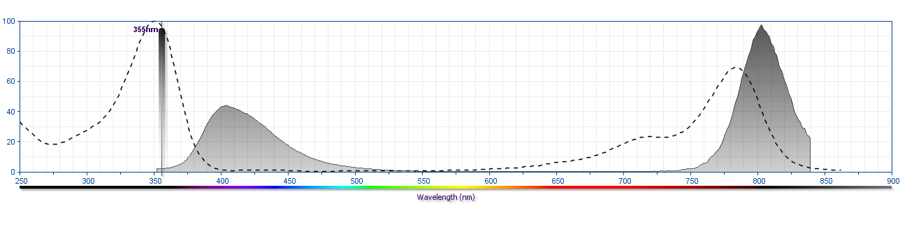
- For fluorochrome spectra and suitable instrument settings, please refer to our Multicolor Flow Cytometry web page at www.bdbiosciences.com/colors.
- An isotype control should be used at the same concentration as the antibody of interest.
- BD Horizon Brilliant Ultraviolet 805 is covered by one or more of the following US patents: 8,110,673, 8,158,444; 8,227,187; 8,575,303; 8,354,239.
- BD Horizon Brilliant Stain Buffer is covered by one or more of the following US patents: 8,110,673; 8,158,444; 8,575,303; 8,354,239.
- Please refer to http://regdocs.bd.com to access safety data sheets (SDS).
- Human donor specific background has been observed in relation to the presence of anti-polyethylene glycol (PEG) antibodies, developed as a result of certain vaccines containing PEG, including some COVID-19 vaccines. We recommend use of BD Horizon Brilliant™ Stain Buffer in your experiments to help mitigate potential background. For more information visit https://www.bdbiosciences.com/en-us/support/product-notices.
- Species cross-reactivity detected in product development may not have been confirmed on every format and/or application.
Companion Products


The K112-91 monoclonal antibody specifically binds to Bcl-6. Bcl-6 was first identified as a proto-oncogene frequently deregulated by chromosomal translocations in non-Hodgkin B-cell lymphomas. It is a nuclear transcriptional repressor of the BTB/POZ zinc-finger family of transcription factors. In addition to its roles in cancer, Bcl-6 plays important roles in the differentiation of normal cells including B cells, thymocytes, CD4+ or CD8+ T cells. Bcl-6 is highly expressed in germinal center B cells, where it promotes the germinal center reaction by inducing proliferation and inhibiting the DNA-damage response. Bcl-6 has been identified as a key factor in promoting the differentiation of CD4+ follicular T helper (Tfh) cells that are involved in promoting germinal center formation and providing help to B cells. The interplay of Bcl-6 and another transcriptional repressor, Blimp-1, is thought to be critical in defining the results of both B-cell and T-cell differentiation.
Development References (10)
-
Baumjohann D, Okada T, Ansel KM. Cutting Edge: Distinct Waves of BCL6 Expression during T Follicular Helper Cell Development. J Immunol. 2011; 187(5):2089-2092. (Clone-specific: Flow cytometry). View Reference
-
Chung Y, Tanaka S, Chu F, et al. Follicular regulatory T cells expressing Foxp3 and Bcl-6 suppress germinal center reactions. Nat Med. 2011; 17(8):983-988. (Clone-specific: Flow cytometry). View Reference
-
Crotty S, Choi YS, Kageyama R, et al. ICOS receptor instructs T follicular helper cell versus effector cell differentiation via induction of the transcriptional repressor Bcl6. Immunity. 2011; 34:1-15. (Clone-specific: Flow cytometry). View Reference
-
Crotty S, Johnston RJ, Schoenberger SP. Effectors and memories: Bcl-6 and Blimp-1 in T and B lymphocyte differentiation. Nat Immunol. 2010; 11(2):114-120. (Biology). View Reference
-
Crotty S. Follicular Helper CD4 T Cells (Tfh). Annu Rev Immunol. 2011; 29(1):621-663. (Biology). View Reference
-
Eto, D., C. Lao, et al. IL-21 and IL-6 are critical for different aspects of B cell immunity and redundantly induce optimal follicular helper CD4 T cell (Tfh) differentiation. PLoS ONE. 2011; 6(3):e17739. (Clone-specific: Flow cytometry). View Reference
-
Fazilleau N, McHeyzer-Williams LJ, Rosen H, McHeyzer-Williams MG. The function of follicular helper T cells is regulated by the strength of T cell antigen receptor binding. Nat Rev Immunol. 2009; 10(4):375-384. (Biology). View Reference
-
Johnston RJ, Poholek AC, DiToro D, et al. Bcl6 and Blimp-1 are reciprocal and antagonistic regulators of T follicular helper cell differentiation.. Science. 2009; 325(5943):1006-10. (Biology). View Reference
-
Klein U, Dalla-Favera R. Germinal centres: role in B-cell physiology and malignancy. Nat Rev Immunol. 2008; 8(1):22-33. (Biology). View Reference
-
Nurieva RI, Chung Y, Martinez GJ, et al. Bcl6 mediates the development of T follicular helper cells. Science. 2009; 325(5943):1001-1005. (Biology). View Reference
Please refer to Support Documents for Quality Certificates
Global - Refer to manufacturer's instructions for use and related User Manuals and Technical data sheets before using this products as described
Comparisons, where applicable, are made against older BD Technology, manual methods or are general performance claims. Comparisons are not made against non-BD technologies, unless otherwise noted.
For Research Use Only. Not for use in diagnostic or therapeutic procedures.