







Preparation And Storage


Recommended Assay Procedures
Protocol for PBMC cells fixation, permeabilization and staining
1. Harvest, count and pellet PBMC cells following standard procedures.
2. While vortexing, add 5 ml cold 70% - 80% ethanol dropwise into the cell pellet (1-5 x 10^7 cells). Incubate at -20°C for at least 2 hours. These fixed cells can be stored at -20°C for up to 60 days prior to staining.
3. Wash twice with 30-40 ml staining buffer (PBS with 1% FBS, 0.09% NaN3), centrifuge for 10 minutes at 200 x g.
4. Resuspend the cells to a concentration of 1 x 10^7/ml.
5. Transfer 100 µl (1 x 10^6 cells) cell suspension into each sample tube.
6. Add 20 µl of properly diluted fluorescence conjugated antibody into the tubes above. Mix gently.
7. Incubate the tubes at room temperature (RT) for 20-30 minutes in the dark.
8. Wash with 2 ml of staining buffer at 200 x g for 5 minutes.
9. Aspirate the supernatant.
10. Add 0.5 ml of staining buffer to each tube.
11. Proceed to flow cytometric analysis.
Product Notices
- This reagent has been pre-diluted for use at the recommended Volume per Test. We typically use 1 × 10^6 cells in a 100-µl experimental sample (a test).
- An isotype control should be used at the same concentration as the antibody of interest.
- Caution: Sodium azide yields highly toxic hydrazoic acid under acidic conditions. Dilute azide compounds in running water before discarding to avoid accumulation of potentially explosive deposits in plumbing.
- Source of all serum proteins is from USDA inspected abattoirs located in the United States.
- For fluorochrome spectra and suitable instrument settings, please refer to our Multicolor Flow Cytometry web page at www.bdbiosciences.com/colors.
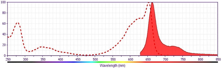
- This APC-conjugated reagent can be used in any flow cytometer equipped with a dye, HeNe, or red diode laser.
- Please refer to http://regdocs.bd.com to access safety data sheets (SDS).
- Please refer to www.bdbiosciences.com/us/s/resources for technical protocols.

Monoclonal antibody APA1/1 reacts with the intracellular domain of the epsilon chain of the CD3 molecule (CD3-ε). This antibody does not react with the extracellular region of CD3-ε. The assembly of the T cell antigen receptor (TCR)-CD3 complex has been suggested to take place by pairwise interactions of the CD3-ε subunit with either CD3-γ or CD3-δ. These dimers then associate with the TCR heterodimer a/β or γ/δ, and the CD3-ζ homodimer to form the full complex. Studies show that antibodies APA1/1 and SP34 gave a strong reaction with COS cells singly transfected with CD3-ε. Other anti-CD3 antibodies (OKT3, WT31, UCHT1, Leu4) did not react with COS cells singly transfected with CD3-ε. This reagent could be useful for the study of T cell development or the study of conformational changes of CD3-ε upon ligand binding to TCR-CD3 complex.
Development References (4)
-
Barroto A, Mallabiabarrena A, Albar JP, Martinez-A C, Alarcon B. Characterization of the region involved in CD3 pairwise interactions within the T cell receptor complex. J Biol Chem. 1998; 273:12807-12816. (Biology).
-
Gil D, Schamel WWA, Montoya M, Sanchez-Madrid F, and Alarcon B. Recruitment of Nck by CD3-epsilon reveals a ligand-induced conformational change essential for T cell receptor signaling and synapse formation. Cell. 2002; 109(7):901-912. (Biology). View Reference
-
Slameron A, Sanchez-Madrid F, Ursa MA, Fresno M, and Alarcon B. A conformational epitope expressed upon association of the CD3-epsilon with either CD3-delta or CD3-gamma is the main target for recognition by anti-CD3 monoclonal antibodies. J Immunol. 1991; 147:3047-3052. (Biology).
-
de la Cruz J, Kruger T, Parks CA, et al. Basal and antigen-induced exposure of the proline-rich sequence in CD3ε.. J Immunol. 2011; 186(4):2282-90. (Clone-specific). View Reference
Please refer to Support Documents for Quality Certificates
Global - Refer to manufacturer's instructions for use and related User Manuals and Technical data sheets before using this products as described
Comparisons, where applicable, are made against older BD Technology, manual methods or are general performance claims. Comparisons are not made against non-BD technologies, unless otherwise noted.
For Research Use Only. Not for use in diagnostic or therapeutic procedures.
