

Preparation And Storage
Product Notices
- Since applications vary, each investigator should titrate the reagent to obtain optimal results.
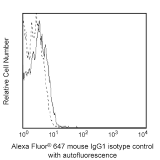
- An isotype control should be used at the same concentration as the antibody of interest.
- Alexa Fluor® 647 fluorochrome emission is collected at the same instrument settings as for allophycocyanin (APC).
- For fluorochrome spectra and suitable instrument settings, please refer to our Multicolor Flow Cytometry web page at www.bdbiosciences.com/colors.
- Caution: Sodium azide yields highly toxic hydrazoic acid under acidic conditions. Dilute azide compounds in running water before discarding to avoid accumulation of potentially explosive deposits in plumbing.
- The Alexa Fluor®, Pacific Blue™, and Cascade Blue® dye antibody conjugates in this product are sold under license from Molecular Probes, Inc. for research use only, excluding use in combination with microarrays, or as analyte specific reagents. The Alexa Fluor® dyes (except for Alexa Fluor® 430), Pacific Blue™ dye, and Cascade Blue® dye are covered by pending and issued patents.
- Alexa Fluor® is a registered trademark of Molecular Probes, Inc., Eugene, OR.
- Please refer to www.bdbiosciences.com/us/s/resources for technical protocols.
Companion Products


The SH1 monoclonal antibody specifically binds to mouse Siglec-G (sialic acid binding immunoglobulin-like lectin G), also known as Siglec10. Siglec-G is a Type I transmembrane glycoprotein that belongs to the Immunoglobulin superfamily. Siglec-G functions as an adhesion molecule that mediates sialic-acid dependent binding to cells. Siglec-G is expressed on cells of the B cell lineage. Siglec-G is most highly expressed by pre-B cells and B1a cells within the B cell lineage and is not detectable on T cells. Siglec-G can inhibit B cell receptor-mediated calcium signaling when it is overexpressed. Mice that lack Siglec-G have significantly increased numbers of B1a cells that begins early in development and is B cell intrinsic. Siglec-G-deficient mice have higher titers of natural IgM antibodies than their normal counterparts. Mouse Siglec-G is the ortholog of human Siglec-10 (also known as, CD330).
Development References (4)
-
Aizawa H, Zimmermann N, Carrigan PE, Lee JJ, Rothenberg ME, Bochner BS. Molecular analysis of human Siglec-8 orthologs relevant to mouse eosinophils: identification of mouse orthologs of Siglec-5 (mSiglec-F) and Siglec-10 (mSiglec-G). Genomics. 2003; 82(5):521-530. (Biology). View Reference
-
Hoffmann A, Kerr S, Jellusova J, et al. Siglec-G is a B1 cell-inhibitory receptor that controls expansion and calcium signaling of the B1 cell population. Nat Immunol. 2007; 8(7):695-704. (Biology). View Reference
-
Jellusova J, Düber S, Gückel E, et al. Siglec-G regulates B1 cell survival and selection. J Immunol. 2010; 185(6):3277-3284. (Biology). View Reference
-
Nitschke L. CD22 and Siglec-G: B-cell inhibitory receptors with distinct functions. Immunol Rev. 2009; 230(1):128-143. (Biology). View Reference
Please refer to Support Documents for Quality Certificates
Global - Refer to manufacturer's instructions for use and related User Manuals and Technical data sheets before using this products as described
Comparisons, where applicable, are made against older BD Technology, manual methods or are general performance claims. Comparisons are not made against non-BD technologies, unless otherwise noted.
For Research Use Only. Not for use in diagnostic or therapeutic procedures.