Old Browser
This page has been recently translated and is available in French now.
Looks like you're visiting us from {countryName}.
Would you like to stay on the current country site or be switched to your country?


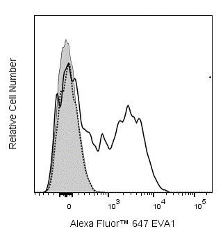
Flow cytometric analysis of EVA1 expression on mouse thymocytes. C57BL/6 mouse thymocytes were stained with PE Anti-Mouse CD4 (Cat. No. 553048/553049/561837), BD Horizon™ BUV737 Rat Anti-Mouse CD8a (Cat. No. 612759), FITC Rat Anti-Mouse CD44 (Cat. No. 553133/561859), BD Horizon™ BUV395 Rat Anti-Mouse CD25 (Cat. No. 564022), and Alexa Fluor™ 647 Mouse IgG1, κ Isotype Control (Cat. No. 565571) or Alexa Fluor™ 647 Mouse Anti-Mouse EVA1 antibody (Cat. No. 567660) at 0.5 μg/test. DAPI (4',6-Diamidino-2-Phenylindole, Dihydrochloride) Solution (Cat. No. 564907) was added to cells right before analysis. Isotype control (dotted line histogram) or EVA1 (solid line histogram) staining on DN3 cells (CD4- CD8a- CD44- CD25+) are shown and compared to EVA1 (shaded histogram) staining on DN1 cells (CD4- CD8a- CD44+ CD25-). The fluorescence histograms showing the expression of EVA1 (or Ig Isotype control staining) were derived from gated events with the forward and side light-scatter characteristics of viable (DAPI-negative) thymocytes. Flow cytometry and data analysis were performed using a BD X-20 LSRFortessa™ Cell Analyzer System and FlowJo™ software. Data shown on this Technical Data Sheet are not lot specific.


BD Pharmingen™ Alexa Fluor™ 647 Mouse Anti-Mouse EVA1

Regulatory Status Legend
Any use of products other than the permitted use without the express written authorization of Becton, Dickinson and Company is strictly prohibited.
Preparation And Storage
Recommended Assay Procedures
BD® CompBeads can be used as surrogates to assess fluorescence spillover (Compensation). When fluorochrome conjugated antibodies are bound to BD® CompBeads, they have spectral properties very similar to cells. However, for some fluorochromes there can be small differences in spectral emissions compared to cells, resulting in spillover values that differ when compared to biological controls. It is strongly recommended that when using a reagent for the first time, users compare the spillover on cells and BD® CompBeads to ensure that BD® CompBeads are appropriate for your specific cellular application.
Product Notices
- Please refer to www.bdbiosciences.com/us/s/resources for technical protocols.
- Alexa Fluor® 647 fluorochrome emission is collected at the same instrument settings as for allophycocyanin (APC).
- Caution: Sodium azide yields highly toxic hydrazoic acid under acidic conditions. Dilute azide compounds in running water before discarding to avoid accumulation of potentially explosive deposits in plumbing.
- Since applications vary, each investigator should titrate the reagent to obtain optimal results.
- For fluorochrome spectra and suitable instrument settings, please refer to our Multicolor Flow Cytometry web page at www.bdbiosciences.com/colors.
- An isotype control should be used at the same concentration as the antibody of interest.
- This product is provided under an intellectual property license between Life Technologies Corporation and BD Businesses. The purchase of this product conveys to the buyer the non-transferable right to use the purchased amount of the product and components of the product in research conducted by the buyer (whether the buyer is an academic or for-profit entity). The buyer cannot sell or otherwise transfer (a) this product (b) its components or (c) materials made using this product or its components to a third party or otherwise use this product or its components or materials made using this product or its components for Commercial Purposes. Commercial Purposes means any activity by a party for consideration and may include, but is not limited to: (1) use of the product or its components in manufacturing; (2) use of the product or its components to provide a service, information, or data; (3) use of the product or its components for therapeutic, diagnostic or prophylactic purposes; or (4) resale of the product or its components, whether or not such product or its components are resold for use in research. For information on purchasing a license to this product for any other use, contact Life Technologies Corporation, Cell Analysis Business Unit Business Development, 29851 Willow Creek Road, Eugene, OR 97402, USA, Tel: (541) 465-8300. Fax: (541) 335-0504.
- Please refer to http://regdocs.bd.com to access safety data sheets (SDS).
- Alexa Fluor™ is a trademark of Life Technologies Corporation.
- For U.S. patents that may apply, see bd.com/patents.
Companion Products


The G9P3 monoclonal antibody specifically recognizes mouse EVA1, which is encoded by the Mpzl2 gene. EVA1 is a member of the immunoglobulin-like cell-adhesion molecule (Ig-CAM or IgSF CAM) family, members of which are anchored to the surface of epithelial and endothelial cells via a GPI domain. This class of adhesion molecules interacts intracellularly with cytoskeletal proteins and extracellularly with Ig-CAM family members, by homophilic or heterophilic binding, and with other classes of adhesion molecules such as integrins and cadherins. EVA1 is expressed on and is involved in the structural integrity of epithelia throughout the body, eg mammary glands, liver, inner ear, and the blood-brain barrier. In the thymus, EVA1 is expressed both on thymic epithelial cells (TECs) and on a subset of developing thymocytes, known as DN3. It is a low-affinity mediator of developmental signals between thymocytes and TEC that support the differentiation and survival of both developing thymocytes and the TECs. G9P3 mAb was produced in EVA1 knockout mice immunized with recombinant mouse EVA1 extracellular region, and it recognizes mouse and human EVA1 expressed on transfected cells.
Development References (10)
-
Chatterjee G, Carrithers LM, Carrithers MD. Epithelial V-like antigen regulates permeability of the blood-CSF barrier.. Biochem Biophys Res Commun. 2008; 372(3):412-7. (Biology). View Reference
-
Garabatos N, Blanco J, Fandos C, et al. A monoclonal antibody against the extracellular domain of mouse and human epithelial V-like antigen 1 reveals a restricted expression pattern among CD4- CD8- thymocytes.. Monoclon Antib Immunodiagn Immunother. 2014; 33(5):305-11. (Immunogen: Flow cytometry). View Reference
-
Guttinger M, Sutti F, Panigada M, et al. Epithelial V-like antigen (EVA), a novel member of the immunoglobulin superfamily, expressed in embryonic epithelia with a potential role as homotypic adhesion molecule in thymus histogenesis.. J Cell Biol. 1998; 141(4):1061-71. (Biology). View Reference
-
Iacovelli S, Iosue I, Di Cesare S, Guttinger M. Lymphoid EVA1 expression is required for DN1-DN3 thymocytes transition. PLoS One. 2009; 4(10):e7586. (Biology). View Reference
-
Kurd N, Robey EA. T-cell selection in the thymus: a spatial and temporal perspective.. Immunol Rev. 2016; 271(1):114-26. (Biology). View Reference
-
Lin X, Cui M, Xu D, et al. Liver-specific deletion of Eva1a/Tmem166 aggravates acute liver injury by impairing autophagy.. Cell Death Dis. 2018; 9(7):768. (Biology). View Reference
-
Litvinov IV, Netchiporouk E, Cordeiro B, et al. Ectopic expression of embryonic stem cell and other developmental genes in cutaneous T-cell lymphoma.. Oncoimmunology. 2014; 3(11):e970025. (Biology). View Reference
-
Ramena G, Yin Y, Yu Y, Walia V, Elble RC. CLCA2 Interactor EVA1 Is Required for Mammary Epithelial Cell Differentiation.. PLoS ONE. 2016; 11(3):e0147489. (Biology). View Reference
-
Wesdorp M, Murillo-Cuesta S, Peters T, et al. MPZL2, Encoding the Epithelial Junctional Protein Myelin Protein Zero-like 2, Is Essential for Hearing in Man and Mouse.. Am J Hum Genet. 2018; 103(1):74-88. (Biology). View Reference
-
Wright E, Rahgozar K, Hallworth N, Lanker S, Carrithers MD. Epithelial V-like antigen mediates efficacy of anti-alpha₄ integrin treatment in a mouse model of multiple sclerosis.. PLoS ONE. 2013; 8(8):e70954. (Biology). View Reference
Please refer to Support Documents for Quality Certificates
Global - Refer to manufacturer's instructions for use and related User Manuals and Technical data sheets before using this products as described
Comparisons, where applicable, are made against older BD Technology, manual methods or are general performance claims. Comparisons are not made against non-BD technologies, unless otherwise noted.
For Research Use Only. Not for use in diagnostic or therapeutic procedures.
Report a Site Issue
This form is intended to help us improve our website experience. For other support, please visit our Contact Us page.