







Preparation And Storage


Product Notices
- This reagent has been pre-diluted for use at the recommended Volume per Test. We typically use 1 × 10^6 cells in a 100-µl experimental sample (a test).
- Please refer to www.bdbiosciences.com/us/s/resources for technical protocols.
- For fluorochrome spectra and suitable instrument settings, please refer to our Multicolor Flow Cytometry web page at www.bdbiosciences.com/colors.
- The Alexa Fluor®, Pacific Blue™, and Cascade Blue® dye antibody conjugates in this product are sold under license from Molecular Probes, Inc. for research use only, excluding use in combination with microarrays, or as analyte specific reagents. The Alexa Fluor® dyes (except for Alexa Fluor® 430), Pacific Blue™ dye, and Cascade Blue® dye are covered by pending and issued patents.
- Alexa Fluor® 647 fluorochrome emission is collected at the same instrument settings as for allophycocyanin (APC).
- Alexa Fluor® is a registered trademark of Molecular Probes, Inc., Eugene, OR.
- Caution: Sodium azide yields highly toxic hydrazoic acid under acidic conditions. Dilute azide compounds in running water before discarding to avoid accumulation of potentially explosive deposits in plumbing.
- Source of all serum proteins is from USDA inspected abattoirs located in the United States.
Companion Products
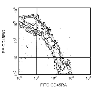
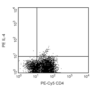
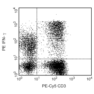
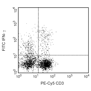
Human IL-17A, also known as IL-17, is a proinflammatory cytokine that is encoded by the IL17A gene in chromosome 6. IL-17A is produced as a disulfide-linked homodimer comprised of two mature 136-amino acid polypeptides. It is a member of the IL-17 family of structurally related cytokines, designated IL-17A through IL-17F. Activated memory T cells, especially Th17 cells (specialized IL-17A-producing CD4+ T cells distinct from Th1 and Th2 cells) produce IL-17 and provide protective immunity against pathogens. Activated CD8+ T cells, γδT cells, NK cells and neutrophils can also be activated to produce IL-17A. IL-17A binds to and exerts its biological activity through IL-17 receptors (IL-17R) that are expressed by a variety of target cells including fibroblasts, epithelial and endothelial cells, monocytes/macrophages and mast cells. The ubiquitous IL-17R expression pattern may explain the broad tissue responsiveness to IL-17. IL-17 induces stromal cells to secrete cytokines and chemokines involved in inflammatory and hematopoietic processes. For example, IL-17 induces fibroblasts to produce IL-6, IL-8, G-CSF and express increased surface ICAM-1. The N49-653 antibody reacts with human IL-17A.
Development References (6)
-
Fossiez F, Djossou O, Chomarat P, et al. T cell interleukin-17 induces stromal cells to produce proinflammatory and hematopoietic cytokines. J Exp Med. 1996; 183(6):2593-2603. (Biology). View Reference
-
Korn T, Oukka M, Kuchroo V, Bettelli E. Th17 cells: effector T cells with inflammatory properties. Semin Immunol. 2007; 19(6):362-371. (Biology). View Reference
-
Moseley TA, Haudenschild DR, Rose L, Reddi AH. Interleukin-17 family and IL-17 receptors. Cytokine Growth Factor Rev. 2003; 14(2):155-174. (Biology). View Reference
-
Weaver CT, Hatton RD, Mangan PR, Harrington LE. IL-17 family cytokines and the expanding diversity of effector T cell lineages. Annu Rev Immunol. 2007; 25:821-852. (Biology). View Reference
-
Yao Z, Painter SL, Fanslow WC, et al. Human IL-17: a novel cytokine derived from T cells. J Immunol. 1995; 155(12):5483-5486. (Immunogen). View Reference
-
Yao Z, Spriggs MK, Derry JM, et al. Molecular characterization of the human interleukin (IL)-17 receptor. Cytokine. 1997; 9(11):794-800. (Biology). View Reference
Please refer to Support Documents for Quality Certificates
Global - Refer to manufacturer's instructions for use and related User Manuals and Technical data sheets before using this products as described
Comparisons, where applicable, are made against older BD Technology, manual methods or are general performance claims. Comparisons are not made against non-BD technologies, unless otherwise noted.
For Research Use Only. Not for use in diagnostic or therapeutic procedures.
