Old Browser
This page has been recently translated and is available in French now.
Looks like you're visiting us from {countryName}.
Would you like to stay on the current country site or be switched to your country?


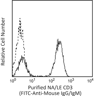
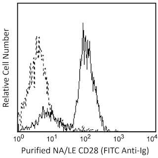
Analysis of CD247 (CD3ζ) (pY142) in activated human T leukemia cells. Jurkat cells (ATCC TIB-152) were either stimulated by cross-linking of CD3 and CD28 with NA/LE Mouse anti-Human CD3 mAb UCHT1 (Cat. No. 555329) and NA/LE Mouse anti-Human CD28 mAb CD28.2 (Cat. No. 555725) on ice for 15 minutes followed by Purified Goat anti-Mouse Ig (Cat. No. 553998) on ice for 15 minutes, and then allowed to undergo phosphorylation at 37°C for 2 minutes (shaded histogram) or unstimulated (open histogram). The cells were fixed (BD Phosflow™ Fix Buffer I, Cat. No. 557870) for 10-15 minutes at 37°C, permeabilized (BD Phosflow™ Perm Buffer III, Cat. No. 558050) on ice for at least 30 minutes, blocked with normal mouse immunoglobulin, and then stained with Alexa Fluor® 647 Mouse anti-CD247 (CD3ζ) (pY142). Flow cytometry was performed on a BD FACSCalibur™ flow cytometry system.


BD™ Phosflow Alexa Fluor® 647 Mouse anti-CD247 (pY142)

Regulatory Status Legend
Any use of products other than the permitted use without the express written authorization of Becton, Dickinson and Company is strictly prohibited.
Preparation And Storage
Product Notices
- This reagent has been pre-diluted for use at the recommended Volume per Test. We typically use 1 × 10^6 cells in a 100-µl experimental sample (a test).
- Alexa Fluor® is a registered trademark of Molecular Probes, Inc., Eugene, OR.
- Alexa Fluor® 647 fluorochrome emission is collected at the same instrument settings as for allophycocyanin (APC).
- The Alexa Fluor®, Pacific Blue™, and Cascade Blue® dye antibody conjugates in this product are sold under license from Molecular Probes, Inc. for research use only, excluding use in combination with microarrays, or as analyte specific reagents. The Alexa Fluor® dyes (except for Alexa Fluor® 430), Pacific Blue™ dye, and Cascade Blue® dye are covered by pending and issued patents.
- Caution: Sodium azide yields highly toxic hydrazoic acid under acidic conditions. Dilute azide compounds in running water before discarding to avoid accumulation of potentially explosive deposits in plumbing.
- Source of all serum proteins is from USDA inspected abattoirs located in the United States.
- For fluorochrome spectra and suitable instrument settings, please refer to our Multicolor Flow Cytometry web page at www.bdbiosciences.com/colors.
- Please refer to www.bdbiosciences.com/us/s/resources for technical protocols.
Companion Products


The T cell receptor (TCR), expressed by thymocytes and T lymphocytes, is a multi-component cell-surface complex responsible for recognizing antigen in the context of MHC molecules. The antigen-specific binding component of the TCR, Ti, is a heterodimer of the variable Ig-like subunits α and β or γ and δ. Ti is non-covalently associated with an invariant set of molecules referred to as the CD3 polypeptides, γ, δ, ε, and ζ. The CD3 ζ polypeptide (CD3ζ) was named CD247 at the 7th Human Leukocyte Differentiation Antigens Workshop. CD3 appears early in thymocyte differentiation and remains expressed on all mature T lymphocytes. After antigen recognition by the TCR, CD3ζ is the primary intracellular signal transducing subunit. It contains three ITAMs (Immunoreceptor Tyrosine-based Activation Motifs), each of which contains a pair of tyrosine residues that are phosphorylated by Lck and Fyn and are required for signal propagation. The molecular weight of CD3ζ is 16 kDa, and it is also observed as 32-kDa homodimers or as heterodimers with the γ chain of Fc receptors. Upon phosphorylation, the CD3ζ monomer undergoes an apparent shift in electrophoretic mobility up to 21 kDa.
The K25-407.69 monoclonal antibody recognizes the phosphorylated tyrosine 142 (pY142) in the third ITAM domain of human CD3ζ (CD247).
Development References (4)
-
Alberola-Ila J, Takaki S, Kerner JD, Perlmutter RM. Differential signaling by lymphocyte antigen receptors. Annu Rev Immunol. 1997; 15:125-154. (Biology). View Reference
-
Ernst DN, Shih CC. CD3 complex. J Biol Regul Homeost Agents. 2000; 14(3):226-229. (Biology). View Reference
-
Kersh EN, Shaw AS, Allen PM. Fidelity of T cell activation through multistep T cell receptor ζ phosphorylation. Science. 1998; 281:572-575. (Biology). View Reference
-
Salomon AR, Ficarro SB, Brill LM, et al. Profiling of tyrosine phosphorylation pathways in human cells using mass spectrometry. Proc Natl Acad Sci U S A. 2003; 100(2):443-448. (Biology). View Reference
Please refer to Support Documents for Quality Certificates
Global - Refer to manufacturer's instructions for use and related User Manuals and Technical data sheets before using this products as described
Comparisons, where applicable, are made against older BD Technology, manual methods or are general performance claims. Comparisons are not made against non-BD technologies, unless otherwise noted.
For Research Use Only. Not for use in diagnostic or therapeutic procedures.
Report a Site Issue
This form is intended to help us improve our website experience. For other support, please visit our Contact Us page.