-
Reagents
- Flow Cytometry Reagents
-
Western Blotting and Molecular Reagents
- Immunoassay Reagents
-
Single-Cell Multiomics Reagents
- BD® OMICS-Guard Sample Preservation Buffer
- BD® AbSeq Assay
- BD® Single-Cell Multiplexing Kit
- BD Rhapsody™ ATAC-Seq Assays
- BD Rhapsody™ Whole Transcriptome Analysis (WTA) Amplification Kit
- BD Rhapsody™ TCR/BCR Next Multiomic Assays
- BD Rhapsody™ Targeted mRNA Kits
- BD Rhapsody™ Accessory Kits
- BD® OMICS-One Protein Panels
-
Functional Assays
-
Microscopy and Imaging Reagents
-
Cell Preparation and Separation Reagents
-
- BD® OMICS-Guard Sample Preservation Buffer
- BD® AbSeq Assay
- BD® Single-Cell Multiplexing Kit
- BD Rhapsody™ ATAC-Seq Assays
- BD Rhapsody™ Whole Transcriptome Analysis (WTA) Amplification Kit
- BD Rhapsody™ TCR/BCR Next Multiomic Assays
- BD Rhapsody™ Targeted mRNA Kits
- BD Rhapsody™ Accessory Kits
- BD® OMICS-One Protein Panels
- Germany (English)
-
Change country/language
Old Browser
This page has been recently translated and is available in French now.
Looks like you're visiting us from United States.
Would you like to stay on the current country site or be switched to your country?
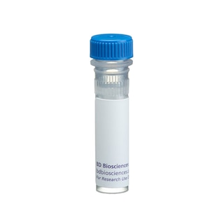
BD Transduction Laboratories™ Purified Mouse Anti-CD29
Clone 18/CD29 (RUO)


Western blot analysis of CD29 (Integrin β1) on a A431 cell lysate (Human epithelial carcinoma; ATCC CRL-1555). Lane 1: 1:2500, lane 2: 1:5000, lane 3: 1: 10,000 diluton of the anti-CD29 antibody.

Immunofluorescence staining of HeLa cells (Human cervical epitheloid carcinoma; ATCC CCL-2.2).


BD Transduction Laboratories™ Purified Mouse Anti-CD29

BD Transduction Laboratories™ Purified Mouse Anti-CD29

Regulatory Status Legend
Any use of products other than the permitted use without the express written authorization of Becton, Dickinson and Company is strictly prohibited.
Preparation And Storage
Recommended Assay Procedures
Western blot: Please refer to http://www.bdbiosciences.com/pharmingen/protocols/Western_Blotting.shtml
Product Notices
- Since applications vary, each investigator should titrate the reagent to obtain optimal results.
- Please refer to www.bdbiosciences.com/us/s/resources for technical protocols.
- Caution: Sodium azide yields highly toxic hydrazoic acid under acidic conditions. Dilute azide compounds in running water before discarding to avoid accumulation of potentially explosive deposits in plumbing.
- Source of all serum proteins is from USDA inspected abattoirs located in the United States.
Data Sheets
Companion Products

.png?imwidth=320)
Integrins are membrane receptors that mediate cell-cell or cell-matrix adhesion. All integrins are transmembrane heterodimers composed of α and β subunits and are connected to the cytoskeleton. At least 20 integrins, formed from combinations of 12 α and 9 β subunits, have been reported. Many of these have been implicated as transducers of molecular signals. The β1 subgroup of this receptor family comprises at least six different dimer combinations. Among these combinations, α2β1 is associated with type I collagen and laminin binding and regulation, α3β1 is a receptor for laminin and epiligrin, and α5β1 is a fibronectin receptor. β1 integrins play an important role in several aspects of epidermal differentiation and morphogenesis. Expression of the β1 subunit is regulated by growth factors such as TGF-β1. Integrin activation, which enhances binding of T cells to endothelium, is regulated in part by phosphatidylinositol 3-kinase. Focal adhesion kinase (FAK) and paxillin have been reported to independently bind the C-terminal, cytoplasmic domain of the β1 subunit. FAK is reported to be enzymatically activated upon engagement of integrins with their ligands and paxillin is phosphorylated on tyrosine upon activation of FAK.
Development References (5)
-
Cervella P, Silengo L, Pastore C, Altruda F. Human beta 1-integrin gene expression is regulated by two promoter regions. J Biol Chem. 1993; 268(7):5148-5155. (Biology). View Reference
-
Huan Y, van Adelsberg J. Polycystin-1, the PKD1 gene product, is in a complex containing E-cadherin and the catenins. J Clin Invest. 1999; 104(10):1459-1468. (Biology: Immunohistochemistry, Western blot). View Reference
-
Ivaska J, Whelan RD, Watson R, Parker PJ. PKC epsilon controls the traffic of beta1 integrins in motile cells. EMBO J. 2002; 21(14):3608-3619. (Biology: Immunofluorescence, Immunoprecipitation, Western blot). View Reference
-
Tang H, Hao Q, Fitzgerald T, Sasaki T, Landon EJ, Inagami T. Pyk2/CAKbeta tyrosine kinase activity-mediated angiogenesis of pulmonary vascular endothelial cells. J Biol Chem. 2002; 277(7):5441-5447. (Biology: Western blot). View Reference
-
Yeh CH, Peng HC, Huang TF. Accutin, a new disintegrin, inhibits angiogenesis in vitro and in vivo by acting as integrin alphavbeta3 antagonist and inducing apoptosis. Blood. 1998; 92(9):3268-3276. (Biology: Flow cytometry). View Reference
Please refer to Support Documents for Quality Certificates
Global - Refer to manufacturer's instructions for use and related User Manuals and Technical data sheets before using this products as described
Comparisons, where applicable, are made against older BD Technology, manual methods or are general performance claims. Comparisons are not made against non-BD technologies, unless otherwise noted.
For Research Use Only. Not for use in diagnostic or therapeutic procedures.