Boost Your Confidence in T-Cell Panels
Introduction to T-cell characterisation
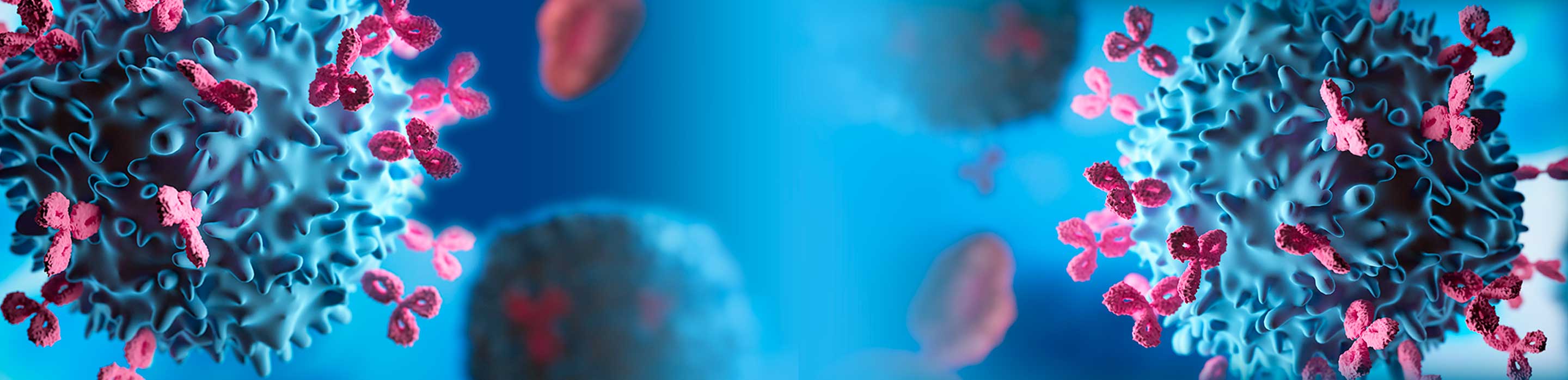
Flow cytometry (FC) is a powerful tool which allows the simultaneous measurement and analysis of multiple phenotypical and functional characteristics of single cells, including CD4+ T helper cell subsets, polyfunctional T cells and regulatory T cells1,2.
Our understanding of the role of T lymphocytes, their application in immune therapy, and their dysregulation in various diseases has been at the forefront of research for the last decade2,3. Researchers are tasked with using more intuitive panels to ensure deeper T-cell characterisation without resolution loss from spillover spread which significantly reduces the sensitivity4.
Challenges facing researchers with existing panels
Time and cost are precious resources for researchers, but timely expansion of existing FC panels is far more complicated than just adding new fluorochromes and markers5. The improvident introduction of multiple florescences causes them to spill into each detector leading to unwanted signals and an overall reduction in sensitivity6.
The spillover spread may also compromise the resolution which limits the ability to define cell biology in great depth5. This ultimately requires a new panel to be completely redesigned, resulting in additional costs and development time5. In-depth T-cell characterisation can also be a challenge using existing FC panels. Characterising sub-sets of T cells using a minimal combination of surface markers is not sufficient for deeper immunophenotypic and functional resolution7,8.
In some cases, entire T-cell subsets may remain undetected or even lost during cell isolation procedures9.
How T-cell panels can help
Larger pre-optimised backbone multicolour panels are the key to ensuring deeper T-cell characterisation. These panels are simple to use and highly accessible5.
Pre-optimised panels are strategically designed to allow the addition of up to five drop-in fluorochromes with minimal panel design effort5. The flexible backbone panels clearly resolve major T-cell subsets without risking spillover spread and resolution loss5.
Application of recent T-cell research
Flow cytometry has applications in multiple disciplines such as immunology, virology, molecular biology, cancer biology, and infectious disease monitoring10. The method has played an instrumental role in our comprehension of the immune system and its interplay with human tumours11.
For immunology studies, flow cytometry is used to observe the immune response to infectious diseases and cancer10. It allows simultaneous differentiation of cells from blood, bone marrow, and solid tissues, as well as characterisation of complex immune phenotypes and flexibility in measuring multiple immune functions10,11.
The technique has recently experienced dramatic advances and the methods and technologies are evolving continuously11. The continued development of more efficient fluorochromes and high technology flow cytometers will lead to significant improvements in medical and biomedical researches1.
Tips and tricks for optimising flow cytometry performance
Follow these helpful tips and painful poor resolutions will be a thing of the past5!
- Perform red blood cell (RBC) lysis post-antibody staining to avoid impact on antigen integrity and antibody binding.
- Perform enrichment via negative selection (magnetic depletion of unwanted cells) to minimise perturbation of cell populations of interest.
- Make sure the signal of the compensation control is as bright or brighter than the experiment sample.
- Do not change fluorescence detector voltage or gain after acquisition of the compensation controls.
- Always test the accuracy of compensation when using cells or compensation beads.