





BD Pharmingen™ Purified Mouse Anti-Insulin
Clone T56-706 (RUO)


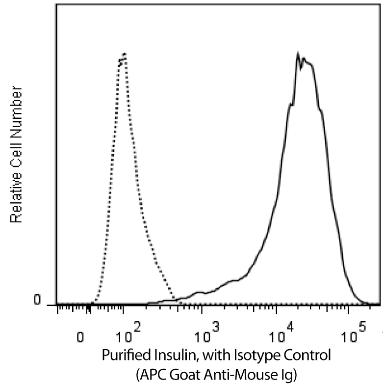
Flow cytometric analysis of insulin expression in human insulin-transfected 293F cells and a mouse insulinoma cell line.
LEFT: Untransfected (dashed line histogram) and human insulin-transfected (solid line histogram) 293F cells were fixed with BD Cytofix™ Fixation Buffer (Cat. No. 554655) and permeabilized with BD Phosflow™ Perm Buffer III (Cat. No. 558050). The cells were then washed and stained with Purified Mouse Anti-Insulin (Cat. No. 565688) followed by APC Goat Anti-Mouse Ig (Cat. No. 550826).
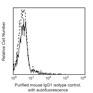
RIGHT: Beta-TC-6 cells (ATCC CRL-11505) were fixed with BD Cytofix™ Fixation Buffer (Cat. No. 554655) and permeabilized with BD Phosflow™ Perm Buffer III (Cat. No. 558050). The cells were then washed and stained with either Purified Mouse IgG1, κ isotype control (Cat. No. 554121), dashed-line histogram) or Purified Mouse Anti-Insulin (Cat. No. 565688, solid line histogram) followed by APC Goat Anti-Mouse Ig (Cat. No. 550826).
All fluorescence histograms were derived from gated events with the forward and side light-scatter characteristics of intact cells. Flow cytometry was performed on a BD Canto™ II flow cytometry system.
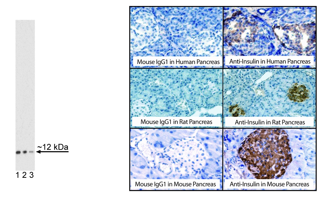
LEFT: Western blot analysis of Insulin expression. Lysate from insulin-transfected 293F cells was prepared for electrophoresis (SDS-PAGE) in a 2D Tris-Glycine polyacrylamide gel. The proteins were transferred to PVDF membranes and then probed with 2.0 (lane 1), 1.0 (lane 2), and 0.5 (lane 3) µg/mL of Purified Mouse Anti-Insulin (Cat. No. 565688). Specific staining was detected with HRP Goat Anti-Mouse Ig (Cat No 554002).
RIGHT: Immunohistochemical staining of insulin in human, rat, and mouse islets of Langerhans. Following antigen retrieval with BD Pharmingen™ Retrievagen A Buffer (Cat. No. 550524), sections from formalin-fixed, paraffin-embedded human (top row), rat (middle row), and mouse (bottom row) pancreata were blocked using an Avidin/ Biotin Blocking Kit (Vector Laboratories, Cat. No. SP-2001) as recommended by the manufacturer. The sections were then stained overnight with either Purified Mouse IgG1 κ Isotype Control (Cat. No. 550878, left column) or Purified Mouse Anti-Insulin (Cat. No. 565688, right column). A three-step staining procedure that employs either Biotin Goat Anti-Mouse Ig (Cat. No. 550337, top and middle rows) or Biotin Rat Anti-Mouse IgG1 (Cat. No.553441, bottom row), Streptavidin HRP (Cat. No. 550946) and DAB (Cat. No. 550880) to develop the primary staining reagents, Counterstaining was with Hematoxylin. Original magnifications: 20X (top and middle rows) and 40X (bottom row).

Flow cytometric analysis of insulin expression in human insulin-transfected 293F cells and a mouse insulinoma cell line.
LEFT: Untransfected (dashed line histogram) and human insulin-transfected (solid line histogram) 293F cells were fixed with BD Cytofix™ Fixation Buffer (Cat. No. 554655) and permeabilized with BD Phosflow™ Perm Buffer III (Cat. No. 558050). The cells were then washed and stained with Purified Mouse Anti-Insulin (Cat. No. 565688) followed by APC Goat Anti-Mouse Ig (Cat. No. 550826).
RIGHT: Beta-TC-6 cells (ATCC CRL-11505) were fixed with BD Cytofix™ Fixation Buffer (Cat. No. 554655) and permeabilized with BD Phosflow™ Perm Buffer III (Cat. No. 558050). The cells were then washed and stained with either Purified Mouse IgG1, κ isotype control (Cat. No. 554121), dashed-line histogram) or Purified Mouse Anti-Insulin (Cat. No. 565688, solid line histogram) followed by APC Goat Anti-Mouse Ig (Cat. No. 550826).
All fluorescence histograms were derived from gated events with the forward and side light-scatter characteristics of intact cells. Flow cytometry was performed on a BD Canto™ II flow cytometry system.
LEFT: Western blot analysis of Insulin expression. Lysate from insulin-transfected 293F cells was prepared for electrophoresis (SDS-PAGE) in a 2D Tris-Glycine polyacrylamide gel. The proteins were transferred to PVDF membranes and then probed with 2.0 (lane 1), 1.0 (lane 2), and 0.5 (lane 3) µg/mL of Purified Mouse Anti-Insulin (Cat. No. 565688). Specific staining was detected with HRP Goat Anti-Mouse Ig (Cat No 554002).
RIGHT: Immunohistochemical staining of insulin in human, rat, and mouse islets of Langerhans. Following antigen retrieval with BD Pharmingen™ Retrievagen A Buffer (Cat. No. 550524), sections from formalin-fixed, paraffin-embedded human (top row), rat (middle row), and mouse (bottom row) pancreata were blocked using an Avidin/ Biotin Blocking Kit (Vector Laboratories, Cat. No. SP-2001) as recommended by the manufacturer. The sections were then stained overnight with either Purified Mouse IgG1 κ Isotype Control (Cat. No. 550878, left column) or Purified Mouse Anti-Insulin (Cat. No. 565688, right column). A three-step staining procedure that employs either Biotin Goat Anti-Mouse Ig (Cat. No. 550337, top and middle rows) or Biotin Rat Anti-Mouse IgG1 (Cat. No.553441, bottom row), Streptavidin HRP (Cat. No. 550946) and DAB (Cat. No. 550880) to develop the primary staining reagents, Counterstaining was with Hematoxylin. Original magnifications: 20X (top and middle rows) and 40X (bottom row).


Preparation And Storage


Product Notices
- Since applications vary, each investigator should titrate the reagent to obtain optimal results.
- Please refer to www.bdbiosciences.com/us/s/resources for technical protocols.
- An isotype control should be used at the same concentration as the antibody of interest.
- Sodium azide is a reversible inhibitor of oxidative metabolism; therefore, antibody preparations containing this preservative agent must not be used in cell cultures nor injected into animals. Sodium azide may be removed by washing stained cells or plate-bound antibody or dialyzing soluble antibody in sodium azide-free buffer. Since endotoxin may also affect the results of functional studies, we recommend the NA/LE (No Azide/Low Endotoxin) antibody format, if available, for in vitro and in vivo use.
- Caution: Sodium azide yields highly toxic hydrazoic acid under acidic conditions. Dilute azide compounds in running water before discarding to avoid accumulation of potentially explosive deposits in plumbing.
- Species cross-reactivity detected in product development may not have been confirmed on every format and/or application.
Companion Products


The T56-706 monoclonal antibody specifically binds to insulin, a member of the insulin family of active peptides. Insulin is an evolutionarily conserved peptide hormone that binds to receptors on target cells (primarily adipose and muscle) to promote the absorption of glucose from the blood, thus regulating fat and carbohydrate metabolism. Insulin is produced by β cells in the islets of Langerhans of the pancreas. There, the precursor molecule, preproinsulin, is cleaved to proinsulin that is in turn cleaved to form the mature insulin hormone, which is composed of two peptides (A- and B-chains) linked by 2 disulfide bonds. Mature insulin is stored in granules in the β cells and is released to the blood in response to metabolic signals such as glucose, the amino acids arginine and leucine, and acetylcholine. Catecholamines can regulate blood glucose levels by either stimulating or inhibiting the release of insulin from β cells. The expression of insulin can be used to monitor the pancreatic differentiation of pluripotent stem cells.
Development References (5)
-
Bell GI, Pictet RL, Rutter WJ, Cordell B, Tischer E, Goodman HM. Sequence of the human insulin gene. Nature. 1980; 284(5751):26-32. (Biology). View Reference
-
D'Amour KA, Bang AG, Eliazer S, et al . Production of pancreatic hormone-expressing endocrine cells from human embryonic stem cells. Nat Biotechnol. 2006; 24(12):1481-1483. (Biology). View Reference
-
Kelly OG, Chan MY, Martinson LA, et al. Cell-surface markers for the isolation of pancreatic cell types derived from human embryonic stem cells. Nat Biotechnol. 2011; 29(8):750-756. (Biology). View Reference
-
Pagliuca FW, Millman JR, Gürtler M, et al. Generation of functional human pancreatic β cells in vitro. Cell. 2014; 159(2):428-439. (Biology). View Reference
-
Rezania A, Bruin JE, Riedel MJ et al. Maturation of human embryonic stem cell-derived pancreatic progenitors into functional islets capable of treating pre-existing diabetes in mice. Diabetes. 2012; 61(8):2016-2029. (Biology). View Reference
Please refer to Support Documents for Quality Certificates
Global - Refer to manufacturer's instructions for use and related User Manuals and Technical data sheets before using this products as described
Comparisons, where applicable, are made against older BD Technology, manual methods or are general performance claims. Comparisons are not made against non-BD technologies, unless otherwise noted.
Please refer to Support Documents for Quality Certificates
Global - Refer to manufacturer's instructions for use and related User Manuals and Technical data sheets before using this products as described
Comparisons, where applicable, are made against older BD Technology, manual methods or are general performance claims. Comparisons are not made against non-BD technologies, unless otherwise noted.
For Research Use Only. Not for use in diagnostic or therapeutic procedures.