Skip to main content Skip to navigation
Single-Cell-Multiomics-Page-Banner

Overview
Down Arrow Up Arrow

Single-cell multiomics (scM) is a cutting-edge approach that promises to revolutionize how we perceive and dissect cellular biology. Multiomics, the integration of genomic, transcriptomic, proteomic and other omics data, might initially seem redundant. Why go through the trouble of integrating multiple layers of molecular information when one omic could theoretically capture it all? The answer lies in the complexity of biological networks. Each omic readout provides valuable insights, but no single “ome” can fully unravel the intricate web of cellular processes. For example, while DNA dictates the potential of a cell, not all genes are transcribed into RNA and not all RNA is translated into proteins. Each criterion provides more information about the overall functioning of the cell.

Why Multiomics Matters Now

Multiomics methods offer more robust classifiers for biological samples, aiding in tasks such as cancer subtype identification and patient stratification. Beyond classification, multiomics provides a deeper understanding of genotype-phenotype relationships, unlocking insights into the fundamental mechanisms underlying biological traits and diseases.


In recognition of its significance, single-cell multimodal omics was named Nature Method of the Year in 2019, underscoring its pivotal role in advancing biological research. As we delve deeper into the realm of single-cell multiomics, we unlock new vistas of understanding, paving the way for breakthroughs in personalized medicine, disease treatment and beyond.


Check out BD tools for single cell-multiomics instrumentsscM reagents and scM informatics analysis solutions.

The Five Omes

Genomics


Genomics is the study of an organism's DNA by deciphering the sequence of nucleotide bases comprising an organism's genome. Researchers gain invaluable insights into genetic variation, heredity and evolutionary relationships.

 
Genomics
performance1

Epigenomics


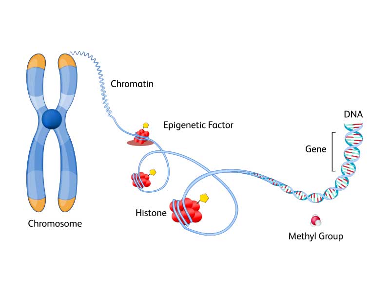
Epigenomics explores the dynamic modifications to DNA and associated proteins that regulate gene expression without altering the underlying genetic code. Epigenetic mechanisms such as DNA methylation, histone modification and non-coding RNA regulation play pivotal roles in orchestrating cellular differentiation, development and response to environmental cues.


Explore BD solutions for single-cell epigenomics.
 
Epigenomics
performance1

Proteomics


Proteomics investigates the entire complement of proteins present within a biological sample. Proteins are the workhorses of the cell, executing diverse functions ranging from catalysis and signaling to structural support and defense. Proteomic analyses offer insights into protein abundance, post- translational modifications and protein-protein interactions.


Explore BD solutions for single-cell proteomics for cell surface proteins and intracellular proteins.

 
performance1

Metabolomics


Metabolomics is the study of small molecule metabolites that serve as the building blocks of cellular metabolism. Metabolites encompass a diverse array of compounds, including amino acids, lipids, sugars and neurotransmitters. Metabolomics elucidates metabolic pathways, metabolic fluxes and metabolic signatures associated with physiological states, environmental exposures and disease phenotypes.


Learn more about the latest trends in single cell multiomics on our blog page.

 
Metabolomics
performance1
Application
Down Arrow Up Arrow

Discover how researchers are leveraging Single-Cell Multiomics in groundbreaking research to advance the field of biology

Immuno-Oncology


  1. Chronic lymphocytic leukemia (CLL)—This study demonstrates the power of single-cell multiomics in unraveling the complexities and heterogeneity of CLL, providing valuable insights for understanding its pathogenesis and potentially informing treatment strategies. Read the datasheet entitled, “Exploring tumor heterogeneity of chronic lymphocytic leukemia using single cell multiomics.”

  2. B cell lymphoma—This study delves into the interplay between malignant B cells and immune cells in B cell lymphoma's tumor microenvironment, offering insights with which to identify therapeutic targets in this disease. Read the datasheet entitled, “Leveraging the power of high-parameter cell sorting and single-cell multiomics to profile intratumoral immune cells in a model of B-cell lymphoma.”

  3. Melanoma—This study investigates tumor induced perturbations in cancer-related hematopoiesis, focusing on how tumors affect different hematopoietic stem and progenitor cell (HSPC) populations. Read the datasheet entitled, “Assessing tumor-driven perturbations during hematopoiesis in a melanoma mouse model through single-cell multiomic analysis.”

  4. Colorectal cancer— A study in Cell Reports Medicine entitled, “Human tumor-infiltrating MAIT cells display hallmarks of bacterial antigen recognition in colorectal cancer” found a specific subset of tumor infiltrating MAIT cells that respond to microbial antigens in colorectal cancer.1 Understanding these cells could pave the way to manipulate them or the microbiome for cancer therapy.
 
Immuno-oncology
performance1

Autoimmunity


  1. Lupus—A study entitled “Chronic immune activation in systemic lupus erythematosus and the autoimmune PTPN22 Trp620 risk allele drive the expansion of FOXP3+ regulatory T cells and PD-1 expression” found that SLE patients have more CD4+FOXP3+ cells due to thymically-derived Tregs expansion linked to PD-1, IL-2, and soluble SIGLEC-11.2

  2. Rheumatoid arthritis (RA)—A Nature Medicine paper entitled “Distinct synovial tissue macrophage subsets regulate inflammation and remission in rheumatoid arthritis” delves into the cellular and molecular mechanisms of sustained remission in RA. It highlights distinct synovial tissue macrophage subsets that regulate inflammation and remission, revealing potential therapeutic targets to encourage resolution of inflammation and restore synovial homeostasis in RA.3

  3. Autoimmune demyelination—A study published in PNAS reveals that TNF-α signaling is crucial in peripheral nerve inflammation and autoimmunity. Aire-deficient mice exhibit increased TNF-α signaling, which recruits and activates immune cells, exacerbating autoimmune demyelination. Blocking TNF-α or its receptors ameliorates neuropathy, suggesting anti-TNF-α therapies could treat inflammatory neuropathies like GBS and CIDP.

  4. Type 1 diabetes mellitus (T1DM)—A study investigated the T cell receptor (TCR) diversity in Japanese patients with T1DM using single-cell VDJ sequencing and showed distinct TCR sequences in autoreactive T cells and differences in gene expression of CD8+ and FOXP3+ cells compared to healthy subjects, suggesting altered TCR diversity and immune response in T1DM.5
 
Autoimmunity
performance1

Infectious Diseases


  1. Acute otitis media—In a rat model of acute otitis media, single-cell transcriptomic profiling revealed dual-feature cells related to phagocytosis. These cells coexisted with professional phagocytes and influenced inflammation progression. Macrophages played a central role in the inflammatory response, with M2 polarization affecting immune microenvironments.6 

  2. COVID-19—A study identifies HIF1A as a long-term immunological scar in monocytes of COVID-19 convalescents without comorbidities. It correlates with disease severity, suggesting its potential as a biomarker for immune monitoring and treatment strategy design post-COVID-19, especially in symptom-free recovery phases.7

  3. Sepsis—Another study investigates mitochondrial STAT3’s role in macrophages during sepsis. It reveals that mitochondrial STAT3 exacerbates sepsis by promoting fatty acid oxidation (FAO) through CPT1a stabilization, mediated by USP501.8 

  4. Tuberculosis-In this study, γδ T cells from a latent tuberculosis donor were enriched and analyzed using single cell immune profiling. It highlights the identification of full-length γδ T cell receptor sequences and clonal expansion, crucial for understanding the immune response to tuberculosis. Read this technote entitled "Single-cell T cell receptor repertoire analysis of sorted γδ T cells using the BD Rhapsody™ TCR/BCR Multiomic Assay".
 
Infectious Diseases
performance1

Immune Cell Biology


  1. Circulating innate lymphoid cells (ILCs)—This study highlights the heterogeneity of ILCs, identified through single-cell multiomic analysis, and the development of high-parameter flow cytometry panels to study ILC subsets across different donors, revealing unique immunophenotypic signatures. Read the datasheet entitled “Resolving human circulating innate lymphoid cell heterogeneity using advanced single cell multiomic analysis.”

  2. Regulatory T cells (Tregs)—This study details the use protein and mRNA single-cell analysis, revealing distinct Treg differentiation states. It showcases the precise resolution of Treg subsets, advancing our understanding of their role in immune homeostasis and disease. Read the datasheet entitled “A comprehensive characterization of regulatory T cells using BD Rhapsody™ Single-Cell Analysis System.”

  3. Neutrophils—This study highlights that neutrophils and emergency granulopoiesis are central to the immune suppression observed in sepsis. Single-cell multiomics revealed distinct populations of immunosuppressive neutrophils and altered granulopoiesis in sepsis patients, suggesting potential targets for therapy and personalized medicine approaches in severe infections.9 

  4. CD8+ T cells—This study describes a novel method called TetTCR-SeqHD, which profiles antigen-specific CD8+ T cells by integrating TCR sequencing with antigen specificity, gene expression and phenotyping. This high-throughput approach offers precise detection of antigen specificity and cross-reactivity, aiding in the study of T cell responses in diseases like type 1 diabetes.10
 
Immune Cell Biology
performance1

Immunotherapy


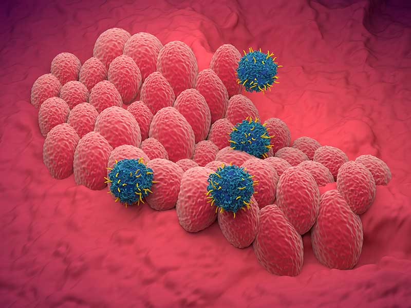
  1. Human head and neck squamous cell carcinomas (HNSCC)—In this study investigating immune alterations in HNSCC and inflamed tissues, researchers found that tumor-unique immune changes exist alongside general inflammatory patterns. Notably, intratumoral IL1R1+ regulatory T (T reg) cells exhibited superior suppressive function compared to IL1R1- T reg cells. Tumor-unique immune alterations, including the role of T reg cells, could have implications for immunotherapy strategies.11 

  2. Lung adenocarcinoma—This study discusses the tumor immune microenvironment (TME) in EGFR mutant lung adenocarcinoma specifically the effect of lower PD-L1 expression and tumor mutation burden (TMB). The study uses single-cell transcriptome analysis to reveal a suppressive TME, with a lack of CD8+ tissue-resident memory cells and insufficient crosstalk between T cells and other cell types.12 

  3. Adoptive T cell therapy (ACT)—This study discusses enhancing ACT for cancer treatment. The researchers found that combining ACT with transient anti-CD4 treatment increases the presence of IL-18Rαhi CD8+ T cells, which improves tumor suppression. This approach could potentially advance immunotherapy research.13
     
  4. Pancreatic ductal adenocarcinoma (PDAC)—This study discusses the role of innate immunity in PDAC, focusing on neutrophils within the tumor microenvironment. It highlights a study revealing various neutrophil subpopulations, including a pro-tumor type associated with poor prognosis. The findings suggest targeting these neutrophils could enhance immunotherapy for PDAC, offering an alternative to T cell-based treatments.14 
 
Immunotherapy
performance1

Neuroscience


  1. Fetal brain development—This study focuses on microglia's role in fetal brain development. It reveals that embryonic microglia accumulate at critical cortical boundaries, preventing cavitary lesions caused by morphogenetic stress. The study highlights microglia's protective functions and the importance of the ATM-factor Spp1 in maintaining structural integrity during brain morphogenesis.15 

  2. Neuropeptide Y (NPY)—The article discusses the role of NPY in regulating immune responses. It highlights how NPY, produced by neurons in the suprarenal and celiac ganglia, can modulate splenic immune activity. The study reveals that NPY influences lymphocyte proliferation and activation, suggesting its potential in treating inflammatory and autoimmune diseases. This neuron-immune system interaction is an ancient mechanism for maintaining immune balance.16 

  3. FGF21 interactions with glutamatergic neurons—This study discusses the hormone FGF21, produced by the liver, and its role in suppressing sugar intake and preference by acting on glutamatergic neurons in the hypothalamus. The study reveals FGF21's direct action in the ventromedial hypothalamus (VMH) affects neuronal activity, specifically regulating sucrose intake without impacting non-nutritive sweet-taste preference, body weight or energy expenditure.17 

  4. Alzheimer's disease (AD)—The article discusses the impact of the ABI3 gene on AD pathology in a mouse model. It reveals that ABI3 deletion increases amyloid β (Aβ) accumulation and impairs microglial function, leading to exacerbated neuroinflammation and synaptic dysfunction. This suggests ABI3's role in AD progression and potential as a therapeutic target.18
 
Neuroscience
performance1
References
  1. Li S, Simoni Y, Becht E, Loh CY, Li N et al., Human tumor-infiltrating MAIT cells display hallmarks of bacterial antigen recognition in colorectal cancer. Cell Rep Med 2020; 23(1)3:100039 doi: 10.1016/j.xcrm.2020.100039
  2. Ferreira R, Dopico XC, Oliveira JJ, Rainbow DB, Yang JH et al., Chronic immune activation in systemic lupus erythematosus and the autoimmune PTPN22 Trp620 risk allele drive the expansion of FOXP3+ regulatory T cells and PD-1 expression” found that SLE patients have more CD4+FOXP3+ cells due to thymically-derived Tregs expansion linked to PD-1, IL-2, and soluble SIGLEC-11. Frontiers in Immunol 2019; 10:2606 https://doi.org/10.3389/fimmu.2019.02606
  3. Alivernini S, MacDonald L, Elmesmari A, Finlay S, Tolusso B, et al. Distinct synovial tissue macrophage subsets regulate inflammation and remission in rheumatoid arthritis. Nat Med 2020; 8:1295-1306. doi: 10.1038/s41591-020-0939-8. Epub 2020 Jun 29
  4. Wang Y, Guo L, Yin X, McCarthy EC, et al. Pathogenic TNF-α drives peripheral nerve inflammation in an Aire-deficient model of autoimmunity. Proc Natl Acad Sci USA 2022; 119(4) e2114406119. doi: 10.1073/pnas.2114406119.
  5. Okamura T, Hamaguchi M, Tominaga H, Kitagawa N, Hoshimoto Y, et al. Characterization of peripheral blood TCR in patients with Type 1 Diabetes Mellitus by BD Rhapsody™ VDJ CDR3 assay. Cells 2022; 11(10):1623. https://doi.org/10.3390/cells11101623
  6. Rao Y, Zhong D, Qiu K, Cheng D, Li L, et al. Single-cell transcriptome profiling identifies phagocytosis-related dual-feature cells in a model of acute otitis media in rats. Front. Immunol. 2021; 12. https://doi.org/10.3389/fimmu.2021.760954
  7. May L, Chu C and Zielinkski CE. Single-Cell RNA Sequencing Reveals HIF1A as a Severity-Sensitive Immunological Scar in Circulating Monocytes of Convalescent Comorbidity-Free COVID-19 Patients. Cells; 2024; 12(4):300. https://doi.org/10.3390/cells13040300
  8. Li R, Li X, Zhao J, Meng F, Yao C, et al. Mitochondrial STAT3 exacerbates LPS-induced sepsis by driving CPT1a-mediated fatty acid oxidation. Theranostics; 2022; 12(2):976-998. doi: 10.7150/thno.63751
  9. Kwok AJ, Allcock A, Ferreira RC, Canco-Gamez E, Smee M, et al. Neutrophils and emergency granulopoiesis drive immune suppression and an extreme response endotype during sepsis. Nat Immunol. 2023; 24: 767-779 https://doi.org/10.1038/s41590-023-01490-5
  10. Ma K, Schonnesen A, He C, Cis AY, Sun E, et al. High-throughput and high-dimensional single-cell analysis of antigen-specific CD8+ T cells. Nat Immunol.; 2021; 22; 1590-1598 https://doi.org/10.1038/s41590-021-01073-2
  11. Mair F, Erickson JR, Frutoso M, Konecny AJ, Greene E, et al. Extricating human tumour immune alterations from tissue inflammation. Nature; 2022; 605(7911):728-735. https://doi.org/10.1038/s41586-022-04718-w
  12. Yang L, He Y-T, Dong S, Wei XX-U, Chen Z-H, et al. Single-cell transcriptome analysis revealed a suppressive tumor immune microenvironment in EGFR mutant lung adenocarcinoma. J Immunother Cancer; 2022; 10(2):e003534.
  13. Kim S, Cho E, Kim Y, Han C, Choi BK, et al. Adoptive immunotherapy with transient anti-CD4 treatment enhances anti-tumor response by increasing IL-18Rαhi CD8+ T cells. Nature Comm; 2022;  5314 https://doi.org/10.1038/s41467-021-25559-7
  14. Wang L, Liu Y, Dai Y, Tang X, Tong Y, et al. Single-cell RNA-seq analysis reveals BHLHE40-driven pro-tumour neutrophils with hyperactivated glycolysis in pancreatic tumour microenvironment. Gut; 2023; 72(5):958-971.
  15. Lawrence A, Canzi A, Bridlance C, Olivie N, Lansonneur C, et al. Microglia maintain structural integrity during fetal brain morphogenesis. Cell; 2024; 187(4):962-980.e19. https://doi.org/10.1016/j.cell.2024.01.012
  16. Yu J, Xiao K, Chen X, Hu J, Fu Z, et al. Neuron-derived neuropeptide Y fine-tunes the splenic immune responses. Neuron; 2022; 110(8):P1327-1339. https://doi.org/10.1016/j.neuron.2022.01.010
  17. Jensen_cody S, Flippo KH, Claflin KE, Yavuz Y, Sapouckey SA, et al. FGF21 Signals to Glutamatergic Neurons in the Ventromedial Hypothalamus to Suppress Carbohydrate Intake. Cell Metab.; 2020; 32(2):273-286. E6. Doi: 10.1016/j.cmet.2020.06.008
  18. Karahan H, Smith DC, Kim B, Dabin LC, Manun Al-Amin Md, et al. Deletion of Abi3 gene locus exacerbates neuropathological features of Alzheimer’s disease in a mouse model of Aβ amyloidosis. Sci Adv. ; 2021; 7(45):eabe3954. doi: 10.1126/sciadv.abe3954
performance1
Plan Your Research
Down Arrow Up Arrow

Whether you're a seasoned researcher or a newcomer to the field, embarking on a single-cell multiomics experiment requires careful planning, meticulous execution and a keen appreciation for the intricacies of multiomic analysis.

performance1

Research Question and Experimental Design

The first step is to clearly define your research question or hypothesis. What specific aspect of cellular biology or disease pathology are you interested in exploring with single-cell multiomics? Are you aiming to identify novel cell types, characterize cellular states or elucidate molecular mechanisms? How many samples/cells do you need to profile to arrive at your desired outcome? To what sequencing depth should you sequence each cell? For example, to characterize subpopulations within a sample you need fewer cells; however, to detect rare cells or do lineage tracing, you need more cells. By articulating your research objectives upfront, you can tailor your experimental design and data analysis strategies to address your scientific goals effectively.


The Satija lab website provides a tool to estimate how many cells you need to sequence so that you see at least “n” number of cells of each type.1

performance1

Select Your Experimental Platform

Single-cell multiomics experiments typically involve the integration of multiple omics modalities, such as genomics, transcriptomics, epigenomics, proteomics and metabolomics. You may also have a choice of different technologies to choose from, each having its own pros and cons. Depending on your research question and available resources, you'll need to select the appropriate experimental platforms and technologies for each omics layer. Consider factors such as sensitivity, throughput, cost, hands on time and compatibility with downstream analysis tools when choosing your experimental platform.


The BD Rhapsody™ HT Single-Cell Analysis System offers a microwell-based single-cell partitioning system coupled with a visual workflow QC system.



Single-cell Multiomics Video playIcon

Sample Preparation and Preservation

Depending on your experimental design, you may need to isolate single cells from tissues, cell cultures or clinical research samples. You may choose manual or automated cell isolation techniques. You may need to enrich or clean up your sample using fluorescence-activated cell sorting (FACS), microfluidic cell sorting, or simple wash and filter. You may also need to preserve your samples using techniques such as cryopreservation, 4 °C preservation and fixation. Careful attention to sample preparation protocols and quality control measures is essential to ensure the reproducibility and accuracy of your results.


Learn more about sample preparation for single-cell multiomics.

performance1

Data Analysis and Integration

The analysis of single-cell multiomics data presents unique computational challenges due to the complexity and dimensionality of the datasets involved. Depending on your expertise and resources, you may choose to perform bioinformatics analysis in-house or collaborate with a commercially available computational platform. Key steps in data analysis include quality control, normalization, dimensionality reduction, clustering and integration of omics datasets to identify biological insights and patterns.


Check out the BD Rhapsody™ Sequence Analysis Pipeline tools and find useful resources on our data analysis page.

performance1

Interpretation and Validation

Once you've analyzed your single-cell multiomics data, it's time to interpret your findings in the context of your research question and existing literature. What biological insights have you gained from your analysis? Do your results support or challenge existing hypotheses? It's essential to critically evaluate your findings and consider alternative explanations before drawing conclusions. Additionally, experimental validation using orthogonal techniques, such as spatial multiomics, qPCR, FACS or functional assays, can help validate key findings and strengthen the robustness of your results.


Learn more about how to process, normalize, interpret, and validate your single-cell multiomics data on our data analysis blog.

performance1
References
  1. How many cells do we need to sample so that we see at least n cells of each type? Satija Lab website; accessed July 19, 2024.
performance1

Overview
Down Arrow Up Arrow


The ability to analyze the expression of genes and proteins at the single-cell level via single-cell multiomics has now enabled deeper insights about cell behavior and function. Single-cell multiomics is now providing users the ability to analyze transcripts and proteins simultaneously across thousands of cells at the single-cell level in the same workflow. BD Biosciences’ multiomic tool, the BD Rhapsody™ Platform and its associated reagents, are all designed to facilitate and support your single-cell multiomic research. 


BD Biosciences tools for single-cell multiomic analyses

The BD Rhapsody™ Single-Cell Analysis System enables high-throughput single-cell multiomic analyses and includes a microwell-based instrument platform equipped with quality monitoring features; an array of RNA and protein assays such as BD Rhapsody™ Whole Transcriptome AssayBD Rhapsody™ Targeted mRNA Panels and BD Rhapsody™ TCR/BCR Profiling AssaysBD® AbSeq Antibody-Oligo’s; and software tools. The BD Rhapsody™ System works seamlessly with other upstream BD cell enrichment technologies such as fluorescence-activated cell sorting enabling users to deep dive into their select cells of interest. Applications for BD single-cell multiomic technology covers a wide spectra of research areas, ranging from studies in immunology and immuno-oncology to understanding the molecular basis of metabolic diseases. The BD Rhapsody™ Platform is paving the way for researchers to apply multiomic tools to dive deep into their respective areas of research. The sections below highlight selected, key application data generated using the BD Rhapsody™ Platform.

 
performance1
Immuno-Oncology
Down Arrow Up Arrow

Immuno-Oncology Research

Chronic Lymphocytic Leukemia Phenotyping

Chronic lymphocytic leukemia (CLL) is the most common form of leukemia in adults and is characterized by a progressive accumulation of functionally incompetent lymphocytes in blood and bone marrow. Recent bulk RNA sequencing studies suggest that there is heterogeneity within CLL samples, demonstrating that single-cell RNA sequencing is necessary to characterize the underlying transcriptional profile. Here is an example of how the BD Rhapsody™ Single-Cell Analysis System was used to simultaneously analyze gene and protein expression in single cells from clinical CLL samples. 


Experimental Outline

(Right) Peripheral blood mononuclear cells (PBMCs) from a total of three healthy donors and four CLL samples were simultaneously stained with BD® Single-Cell Multiplexing Kit reagents, fluorochrome-labeled antibodies and 33 BD® AbSeq Markers. CD19+ cells from different samples were then sorted using the BD FACSMelody™ Cell Sorter and pooled together prior to being loaded on the BD Rhapsody™ System. cDNA was synthesized and processed for library preparation using the targeted BD Rhapsody™ Immune Response Panel (399 genes) before sequencing. Data were analyzed using the BD Rhapsody™ Analysis Pipeline and SeqGeq™ v1.6 Software. 

 
performance1

BD Fluorochrome-Labeled Antibodies Used for Sorting


MarkerFluorochrome
CD3APC
CD14APC
CD16APC
CD19
PE
CD41a
APC
CD56
APC
CD235aAPC

List of fluorochrome-conjugated antibodies (above) and BD® AbSeq Antibodies (right) used in this study.


AbSeq Marker
CD4           
CD24          CD80        CD184        
CD5
CD27CD81CD275
CD7CD33CD86CD279
CD8
CD34CD90CD294
CD9
CD38CD103IgG
CD10
CD40CD117IgD
CD11c
CD45CD123 
CD19CD45RACD127 
CD20CD69CD133 

Multiomic analyses of healthy versus disease samples

To analyze the data generated from the above described workflow, t-SNE analysis was performed on four CLL samples and three healthy donor samples using mRNA expression, surface protein expression or a combination of both. The t-SNE plot generated based on AbSeq data showed higher level of separation among different samples compared to using RNA-Seq data alone. A combination of both mRNA and AbSeq data greatly improved the resolution of different samples. Four CLL samples formed distinct clusters individually whereas the healthy samples overlapped to a greater extent in the t-SNE plot, suggesting the clinical course of CLL patients can be heterogeneous. These results also highlight the power of multiomics in analyzing single-cell profiles.

 
performance1

Differences in key protein markers identified by AbSeq analyses 

CLL is characterized by clonal expansion and accumulation of CD5+ B lymphocytes. Additionally, malignant B cells in CLL patients can also express CD11c at higher levels. BD® AbSeq Reagents revealed higher expression of CD11c and CD5 in CLL samples compared to healthy donors, correlating to pathogenesis of CLL. On the other hand, low CD10 and CD20 expression were observed in CLL samples compared to healthy donors, which is consistent with published literature.

 
performance1

Analysis of differentially expressed markers across healthy and CLL samples

To understand the markers underlying the distinct clustering between healthy and CLL samples, a differential expression analysis was performed using single-cell mRNA and AbSeq data. This analysis identified markers that were decreased in CLL samples compared to healthy samples (Group 1). Genes or proteins that were differentially increased in all CLL samples are listed in Group 2 (all CLL), while Group 3 lists those which are up in some CLL samples. CD5, the marker aberrantly expressed in CLL patients, was higher in CLL03 and CLL04 compared to other samples. Also, a subset of markers that were specifically associated with CLL04 (Group 4) was also discovered, such as CD11c. Interestingly, not all cells produce CD11c at the same level, suggesting intra-tumoral heterogeneity of CLL.

 
performance1

Summary

This study was focused on investigating the differences between three healthy and four CLL samples, utilizing ~500 targeted genes and 33 BD® AbSeq Antibodies. Combining both mRNA and AbSeq data clustered the CLL and healthy samples into distinct groups. Unique markers that are differentially expressed in the CLL samples compared to the healthy control samples were identified in this study. Significant heterogeneity was observed across CLL samples, particularly in the CLL04 sample. Overall, single-cell multiomics of CLL samples revealed novel insights about their unique molecular signature. Such insights for cancers like CLL can potentially aid in designing better therapies. For additional details, download the CLL heterogeneity datasheet detailing the above study.

T Cell Research
Down Arrow Up Arrow


Immunology research—T cell biology


BD has championed in developing cutting-edge tools to drive immunology research for decades. We continue to support immunologists in their single-cell multiomics research as well. Read below to learn about the use of the BD Rhapsody™ Single-Cell Analysis System to address key immunology research questions.

T cell exhaustion study

Deeper characterization of exhausted T cells

Understanding the basis of activation and exhaustion in cell types like T cells has now become the foundation for development of immunotherapy. In some of the fast-developing areas of immunotherapy, like CAR-T therapy, this is becoming even more critical. A key step in the clinical production of CAR-T cells is the expansion of engineered T cells. To generate enough cells for a therapeutic product, cells must be chronically stimulated, which raises the risk of inducing T cell exhaustion and reducing therapeutic efficacy. As protocols for T cell expansion are being developed to optimize CAR T cell yield, function and persistence, fundamental questions about the impact of in vitro manipulation on T cell identity are important to understand. Keeping this in mind, the study described below demonstrates how some of these underlying fundamental questions can be addressed by performing single-cell multiomic analysis utilizing a routinely used model of T cell activation. 


Experimental design 

(Right) Magnetically enriched T cells from PBMCs were stimulated for 3 days in the presence of IL-2 and CD3/CD28 Dynabeads™ Magnetic Beads. Chronic stimulation was carried over for an additional 11 days. Transient stimulation was performed by removing the Dynabeads™ Magnetic Beads and culturing the cells in the presence of IL-2 only for an additional 11 days. A total of five samples were collected and frozen—fresh T cells, cells stimulated for 3, 7 and 14 days (chronically stimulated), and cells stimulated for 3 days and rested for 11 days (transiently stimulated).

 
performance1

(Right) Two aliquots were generated from each time point. One aliquot of each sample was thawed and stained with 39 BD® AbSeq Antibodies and BD® Single-Cell Multiplexing Antibodies. The samples were pooled and loaded into two BD Rhapsody™ Cartridges (~3,000 cells/sample). The BD Rhapsody™ T-Cell Targeted Panel was used to assess the mRNA expression level in these samples. The second aliquot of frozen cells was stained with a 12-color flow cytometry panel and analyzed on the BD LSRFortessa™ X-20 Cell Analyzer.

 
performance1


BD® AbSeq Antibodies and flow antibodies used in the current study

The BD® AbSeq Panel identifies 39 proteins that play roles in T cell activation, inhibition/exhaustion, differentiation or chemotaxis. A companion 12-color flow cytometry panel with selected, overlapping specificities was also designed to assess concordance between flow cytometry and AbSeq technology. scRNA-Seq panel includes 400 genes involved in immune response. All data shown here were from CD8+ T cells.


39-Plex BD® AbSeq Panel
CD4CD278 (ICOS)CD103CD94
CD8CD178 (FAS-L)CD30CD95
CD279 (PD-1)CD183 (CXCR3)CD38CD98
CD223 (LAG-3)CD185 (CXCR5)CD39CD2
CD366 (TIM-3)CD194 (CCR4)CD44TCRab
CD272 (BTLA)CD196 (CCR6)CD45RACD154
CD357 (GITR)/td>CD137 (4-1BB)CD49aCD7
CD226 (DNAM-1)CD134 (OX40)CD54CD2
CD152 (CTLA-4)CD27CD62LCD25
CD270 (HVEM)CD28CD69 

List of BD® AbSeq Antibodies (above) and fluorochrome-conjugated antibodies (right) used for this study.

12-Color Flow Cytometry Panel
MarkerFluorochrome
CD4
BUV805
CD8
BUV395
CD279 (PD-1)
PE-Cy7
CD152 (CTLA-4) 
PE
CD223 (LAG-3)BV480
CD366 (TIM-3)
BV711
CD39
BUV737
CD103
APC
CD45RAAPC-H7
CD62LFITC
CD95BV786
CD357 (GITR)BV421

T cell analyses 

Distinct clusters are resolved for each sample and the resolution is equivalent for clustering based on protein only, mRNA only or a combination of both. The transiently stimulated cell clusters (Day 14) appear separately from both fresh T cells and chronically stimulated cells, suggesting a distinct signature, in contrast to previous observation, suggesting that they are phenotypically and functionally similar to fresh cells. Chronically stimulated cells (Day 3, 7 and 14) form clusters that are separate from fresh cells. The three samples form distinct clusters, suggesting they each have unique signatures.

 
performance1

Analyzing correlation between protein and gene expression at a single-cell level

Analyzing the protein and mRNA signatures of the different populations revealed several insights. Some of these include markers like CD7 where the protein and mRNA expression displayed strong correlation throughout the different time points. CD103 (ITGAE) mRNA was upregulated at Day 3 of activation, while protein expression increases later, at Day 7. In chronically stimulated cells, CD38 mRNA was upregulated at Day 3 and increases over time, whereas protein expression increases later, at Day 14. In transiently stimulated cells, CD38 protein was expressed at Day 14 even though mRNA expression was not maintained.

 
performance1
Concordance of BD® AbSeq Marker data with flow cytometry

The kinetics of protein expression as demonstrated by the percent of CD8 T cell expression markers and the respective level of these cell surface markers detected by both flow cytometry and AbSeq was consistent. These data strongly demonstrate the concordance and utility of both these technologies to study protein expression.

 
performance1

Summary

Use of AbSeq allowed an extensive analysis of a high number of proteins (39 in this study) in a simplified workflow. AbSeq technology enabled resolution of different classes of antigens (primary, secondary, tertiary) that are expressed at various densities (from low to high) on subsets of cells present at different frequencies (from rare to abundant). Additionally, using BD Rhapsody™ System reagents, this study detected changes in expression of both genes and proteins concomitantly over time in different stimulation conditions. Simultaneous analysis of protein and mRNA expression allowed assessment of correlation of mRNA and protein expression to determine potential mechanisms of regulation, leading to a more refined and deeper cell characterization. For additional details, download the T cell exhaustion datasheet detailing the above study.

Regulatory T cell study

Comprehensive characterization of regulatory T cells via single-cell multiomic analysis

Regulatory T cells (Tregs) play an essential role in maintaining immune homeostasis and their alterations can have many implications including tumor progression or onset of autoimmune diseases. Tregs are not a functionally homogenous population and can be subgrouped based on the expression of CD45RA and HLA-DR as naïve or activated Tregs, respectively. This study utilized the BD Rhapsody™ Human Immune Response Panel and BD® AbSeq Antibodies to further dissect Treg subsets and identify putative cell differentiation trajectories. Twenty-two proteins including stimulatory/inhibitory receptors and cytokine/chemokine receptors and 399 mRNA transcripts were analyzed at the single-cell level.


Experimental design 

(Right) Peripheral blood mononuclear cells (PBMCs) from a healthy donor were co-stained with fluorochrome-conjugated antibodies for cell sorting and BD® AbSeq Antibodies for downstream sequencing-based protein detection. The sample was sorted for CD4+CD25+CD127low/− Tregs, and 5,000 sorted Tregs were loaded onto the BD Rhapsody™ Single-Cell Analysis System for single-cell capture. After sample retrieval, BD® AbSeq and mRNA (BD Rhapsody™ Human Immune Response Panel) libraries were prepared for sequencing. The sequencing results were analyzed with the BD Rhapsody™ Analysis Pipeline and SeqGeq™ Software. 

 
performance1

Phenotypic characterization of Tregs cells with BD® AbSeq Antibodies

In this study, the ability of BD® AbSeq Antibodies to clearly identify major Treg subsets previously defined using other technologies was first assessed. The result shows that BD® AbSeq Antibodies enabled a clear resolution of antigens expressed over a continuum such as CD45RA and HLA-DR, which define naïve and activated Tregs, respectively. The expression of selected markers was measured within the CD45RA+HLA-DR naïve cells, CD45RAHLA-DR+ activated cells and CD45RAHLA-DR cells. In agreement with previous reports, CD31 was exclusively detected in a small subset of CD45RA+HLA-DR naïve Tregs, representing recent thymic emigrants (RTEs). On the other hand, CD95 was expressed in both CD45RAHLA-DR and CD45RAHLA-DR+ activated populations, consistent with previous report that identified CD95+ apoptosis-sensitive cells within activated Tregs. Distinct expression patterns were also observed for the chemokine receptor CD185 (CXCR5) and the inhibitory receptor CD279 (PD-1).

 
performance1
Characterization of Treg differentiation by Monocle analysis

Unsupervised high-dimensional data analysis was used to refine the identification of Treg subsets. The Monocle algorithm was used to perform single-cell trajectory analysis and define transitional states of differentiation processes. Based on the expression of 22 protein and 399 mRNA targets, eight distinct states were identified, likely representing distinct differentiation states of Tregs. The single-cell heatmap in the figure below (B) displays selected proteins and genes differentially expressed across the eight states. Cells belonging to each state were arranged, from left to right, based on progressive loss of CD45RA and upregulation of HLA-DR, to represent a progression from naïve to activated cells. In addition to the differential levels of CD45RA expression, these three states could be further discriminated because CD31 is exclusively expressed in state 7 cells, consistent with the signatures of RTEs. On the other hand, the exclusive expression of CD95 and concurrent downregulation of CD45RA in state 5 may suggest a transition from naïve to apoptosis-sensitive and activated cells.

 
performance1
Summary

This study demonstrates the sensitivity and specificity of the BD Rhapsody™ Platform and its multiomic tools, e.g., BD® AbSeq Antibodies, for the identification of Treg subsets based on known surface marker signatures. The combination of single-cell multiomics and high-dimensional data analysis further enabled a clear resolution of distinct subsets of Tregs defined by unique protein and gene expression signatures, likely representing unique differentiation states of Tregs.


For additional details download the regulatory T cells datasheet detailing the above study.

Innate Immune System
Down Arrow Up Arrow

Immunology research—innate immune system


Innate lymphoid cells (ILC) 

Resolving human circulating innate lymphoid cell heterogeneity using single-cell multiomic analysis 

Innate lymphoid cells (ILCs) constitute a recently identified family of lymphoid cells that do not express rearranged antigen receptors and have key functions in inflammation, infection, cancer, metabolic disorders and allergies. They represent a heterogeneous population including classically defined natural killer (NK) cells as well as more recently described non-cytotoxic ILCs. Three major groups of non-cytotoxic ILCs have been characterized in mucosal and non-mucosal tissues based on distinct patterns of cytokine production and transcription factor expression. However, recent publications using single-cell RNA sequencing (scRNA-Seq) or high-parameter mass cytometry showed the existence of more than just three groups of ILCs; hence better understanding of ILC heterogeneity is needed to determine their roles in disease pathogenesis and to determine whether targeting of ILCs will help to prevent or treat these diseases. The emergence of single-cell multiomic technologies provides an opportunity to further resolve ILC heterogeneity by enabling integration of phenotypic and transcriptional data from the same cell.

Experimental outline

Peripheral blood leukocytes were isolated using HetaSep™ (STEMCELL Technologies) and labeled with a panel of 12 fluorochrome-conjugated antibodies for cell sorting. Cells were also simultaneously stained with 42 oligonucleotide-conjugated BD® AbSeq Antibodies for downstream protein detection via next-generation sequencing. A BD Horizon Brilliant Violet™ 510 (BV510) Dye-labeled cocktail of CD19, CD14, TCRγδ, TCRαβ, CD123, CD1a, BDCA2 and FCεR1α antibodies was used as a “dump channel” to exclude lineage cells. To ensure clear resolution and exclusion of T and NK cells, CD3 and CD56 antibodies were additionally included in the sorting panel. Total ILCs were defined as Lineage-CD45+CD127+ cells and were sorted using a BD FACSAria™ Fusion Cell Sorter and loaded into the BD Rhapsody™ Single-Cell Analysis System for single-cell capture. After sample retrieval, single-cell BD® AbSeq and mRNA (BD Rhapsody™ Human Immune Response Panel) libraries were prepared for sequencing. Data were analyzed using the BD Rhapsody™ Analysis Pipeline and SeqGeq™ Software. Biological information discovered using single-cell analyses was then utilized to guide the design of a high-parameter flow cytometry panel. The information derived from multiomic analysis was validated by analyzing peripheral blood ILCs from four different healthy donors using a 21-color flow cytometry panel on the BD FACSymphony™ A5 Cell Analyzer.

 
performance1


MarkerCloneFluorochrome

CD19

CD14

TCRγδ

TCRαβ

CD1a

CD123

BDCA2

FCεR1α

SJ25-C1

MϕP9

11F2

IP26

HI149

9F5

V24-785

AER-37

BV510
CD452D1FITC
CD3SK7APC-H7
CD56B159PE-Cy7
CD127HIL-7R-M21BV421

List of fluorochrome-conjugated antibodies used for sorting Lineage−CD45+CD3CD56CD127+ total ILCs 


BD® AbSeq Panel

MarkerClone
CD2RPA-2.10
CD3SK7
CD4SK3
CD5UCHT2
CD7M-T701
CD8RPA-T8
CD11bM1/70
CD11cB-LY6
CD14MϕP9
CD163G8
CD19SJ25C1
CD252A3
CD27M-T271
CD28CD28.2
CD34581
CD45RAHI100
CD49aSR84
CD49d9F10
CD56NCAM16.2
CD62LDREG-56
CD69FN50
MarkerClone
CD905E10
CD94HP-3D9
CD98UM7F8
CD103BER-ACT8
CD117YB5.B8
CD125 (CTLA-4)BNI3
CD161DX12
CD183 (CXCR3) 1C6/CXCR3
CD184 (CXCR4)12G5
CD196 (CCR6)1149
CD223 (LAG-3) T47-530
CD226 (DNAM-1)DX11
CD274 (PD-L1)MIHI
CD275 (B7-H22D3/B7
CD278 (ICOS)DX29
CD279 (PD-1)EH12.1
CD294 (CRTH2)BM16
CD314 (NKG2D)1D11
CD335 (NKp46)9E2/NKP46
CD336 (NKp44)p44-8
CD366 (TIM-3)7D3

List of BD® AbSeq Oligonucleotide-Conjugated Antibodies used for single-cell multiomic analysis of sorted total ILCs.


Differences in major ILC subgroups revealed by multiomic analyses

Data analysis was initially performed by manually gating the three major groups of circulating ILCs conventionally defined as CD117− CD294− ILC1, CD117+/−CD294+ ILC2 and CD117+CD294− ILC progenitors/ILC3 (ILCP/ILC3). A differential expression analysis was performed between the three ILC groups using single-cell AbSeq and mRNA data and identified unique protein and mRNA signatures for each group. This analysis also revealed expected expression patterns for key transcription factors defining ILC groups. For example, the gene encoding T-bet (TBX21) was exclusively expressed in a subset of ILC1 cells, whereas GATA3 was expressed at highest levels in ILC2 cells. As recently reported, RORC, encoding RORγδ, was partially expressed by circulating ILC progenitors displaying an ILC3 phenotype (CD117+CD294− ILCP/ ILC 3 cells).

 
performance1

Insights about the heterogeneity of ILC1 population

The single-cell heatmap also revealed heterogeneity within each ILC group as most of the represented markers were not uniformly expressed by all the cells. These data therefore suggest that additional subsets exist within each group of ILCs. For example, CD4 was exclusively expressed by the majority, but not all, ILC1 cells. This observation agrees with previous reports and was further confirmed by bi-variate plot analysis clearly showing discrete subsets of CD4+ and CD8+ cells within ILC1 but not ILC2 or ILCP/ILC3 cells.

 
performance1

Single-cell multiomic analysis of ILC1 heterogeneity

To further investigate the heterogeneity of ILC1 cells, a differential gene and protein expression analysis was performed between the three subsets of CD4+CD8, CD4CD8+ and CD4CD8 ILC1 populations. Figure B (above) shows a single-cell heatmap displaying representative protein and gene signatures associated with each subset and illustrates the high heterogeneity and complexity of the ILC1 subset. These data demonstrate the power of a single-cell multiomic approach to screen a high number of proteins and genes and discover critical markers required to define and discriminate unique cell subsets within highly heterogeneous samples.


Of the 42 AbSeq markers tested, this study identified 13 surface markers differentially expressed between the three subsets of ILC1. Also, one differentially expressed gene (CD63) coding for the surface protein CD63 was identified. Further, a 21-color flow cytometry panel was designed based on the learnings from the multiomic data generated to rapidly profile multiple samples for ILC’s (data not shown). 

Summary

In this study, a complete workflow for deep characterization of circulating human ILCs was established. Cell sorting enabled the purification of total ILCs for simultaneous downstream analyses of the expression of 42 proteins and 399 genes. Single-cell multiomic analysis revealed complex immunophenotypic and molecular signatures refining the identity of the three main groups of ILCs. The analysis also revealed heterogeneity within ILC1 cells leading to the discovery of markers defining distinct ILC1 cell populations. The biological information provided by the single-cell multiomic screening was instrumental for the design of a high-parameter flow cytometry panel that could be used to deeply characterize ILC1 cells from multiple donors. For additional details, download the ILC datasheet detailing the above study.

Metabolic Disease Research
Down Arrow Up Arrow

Metabolic disease research


Interrogating immune cells signature in mouse models of obesity using single-cell multiomics

Obesity is a major risk factor for the development of various diseases such as type-2 diabetes, cardiovascular disease and cancer. The transition from a simple adiposity to a disease state correlates with an increase in local and systemic inflammation and is also marked by alterations in the number and function of immune cells. Macrophages are an important source of adipose tissue inflammation and several changes in T cell populations are also involved with the development of obesity in animal models. Interrogating these changes through an integrated analysis of mRNA and protein expression at the single-cell level, can offer deeper insights about how high-fat diet (HFD) can induce inflammation and ultimately drive obesity-related disorders. Here is an example of how BD Biosciences flow cytometry and single-cell analysis tools can be used to interrogate immune cells using mouse models of obesity.

Experimental outline

(Right) Thirty proteins and 399 targeted mRNAs were analyzed in this study in approximately 20,000 single cells derived from multiple tissues of mice that received different diet regimens. Through analysis of cell differentiation and activation markers, numerous cell compartments and observed distinct alterations associated with HFD were examined. The HFD and control diet mice were obtained from Jackson Laboratories. Seven-week-old mice were placed on either a control diet or HFD for 17 weeks. Single-cell suspensions were prepared from bone marrow, thymus, spleen and epididymal adipose tissue. Following tissue collection and dissociation, the eight samples indicated in the figure were stained with one of the 12 unique DNA-barcoded antibodies from the BD® Mouse Immune Single-Cell Multiplexing Kit and 30 BD® AbSeq Mouse Antibodies. BD Pharmingen™ FITC Rat Anti-Mouse CD45 Antibody was included in the staining of adipose tissue cells and CD45+ cells were sorted on the BD FACSAria™ Fusion Cell Sorter. The eight samples were pooled and loaded onto the BD Rhapsody™ Single-Cell Analysis System. After sample retrieval, BD® AbSeq, Sample Tag and mRNA (BD Rhapsody™ Mouse Immune Response Panel) libraries were prepared for sequencing. The sequencing results were analyzed with the BD Rhapsody™ Analysis Pipeline and SeqGeq™ Software.

 
performance1

A. Experimental overview and the BD Rhapsody™ System workflow. B. List of BD® AbSeq Antibodies used in this study.

Unbiased cell clustering identifies multiple cell phenotypes across different samples 

t-SNE was generated using the multiomic data generated from the above experiment for data visualization and an unbiased clustering algorithm PhenoGraph, SeqGeq™ Software plug-in, for detection of cell populations. PhenoGraph identified 12 cell clusters across the four different tissue types. Of the 12 clusters, four showed differences in cell density and distribution in the adipose tissue of HFD mouse compared to control clusters 1, 5, 7 and 12. 

 
performance1

Single-cell protein and targeted mRNA-Seq analyses of CD8+ T cells in adipose tissue from control and HFD mice

(Right) Characterization of cluster 12 revealed a high expression level of CD8b on the surface of these cells, suggesting this cluster was primarily comprised of CD8+ T cells. Notably, cluster 12 of the HFD mouse presented a higher concentration of CD8+ T cells than the controls. Differential mRNA and protein expression analyses of this cluster showed an elevated expression of Pdcd1 (CD279, PD-1 mRNA), Lag3 and Tigit, which are putative markers of cells undergoing exhaustion (Panel B).

 
performance1

Monocle analysis of CD8+ T cells from HFD adipose tissue suggests that these cells underwent cell exhaustion

Specific analysis of CD8+ T cells in the adipose tissue using Monocle discriminated five developmental states. The CD8+ T cells from the control mouse belonged to states 1 and 2 whereas cells from the HFD mouse further followed a trajectory toward states 3, 4 and 5. States 2, 3, 4 and 5 from the HFD mouse presented an elevated expression of exhaustion markers such as CD279 (PD-1) protein and mRNA (Pdcd1) as well Tigit.

 
performance1

Summary

These results demonstrate that single-cell analysis performed on the BD Rhapsody™ Single-Cell Analysis System using BD® AbSeq Antibodies and the BD Rhapsody™ Mouse Immune Response Panel provides unique insights about metabolic disorders like obesity. This comprehensive single-cell analysis platform in combination with powerful bioinformatic tools helped unravel unique disease state immune cell profiles including signs of CD8+ T cells exhaustion and other new insights for HFD-induced inflammation.


To learn more, download the obesity-induced inflammation study datasheet detailing the above study

For Research Use Only. Not for use in diagnostic or therapeutic procedures.