Old Browser
This page has been recently translated and is available in French now.
Looks like you're visiting us from {countryName}.
Would you like to stay on the current country site or be switched to your country?


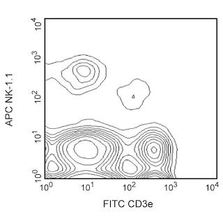
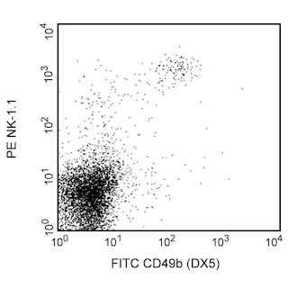
Flow cytometric analysis of CD335 (NKp46) expression on mouse splenocytes. C57BL/6 and BALB/c mouse spleen cells were stained separately with Purified anti-mouse CD335 (NKp46) antibody followed by PE-conjugated Goat anti-rat Ig (Cat. No. 550767). After washing, C57BL/6 cells were stained with APC-conjugated anti-mouse NK-1.1 (NKR-P1B and NKR-P1C) antibody (Cat. No.550627; left panel) and BALB/c cells were stained with FITC-conjugated anti-mouse CD49b (DX5) antibody (Cat. No.553857; right panel). Two-color dot plots showing the correlated expression patterns of CD335/NKp46 and either NK-1.1/CD161 (C57BL/6 cells; left panel) or DX5/CD49b (BALB/c cells; right panel) were derived from gated events with the forward and side light-scatter characteristics of viable lymphocytes. Flow cytometry was performed using a BD™ LSRII System.


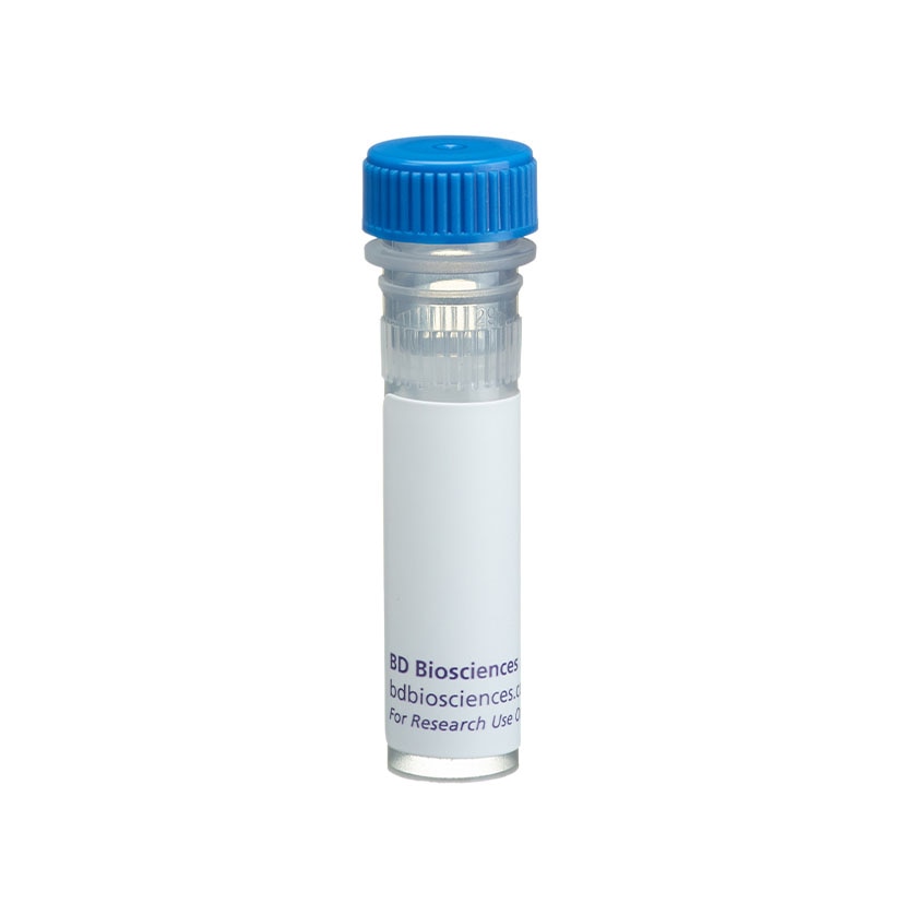
BD Pharmingen™ Purified Rat Anti-Mouse CD335 (NKp46)

Regulatory Status Legend
Any use of products other than the permitted use without the express written authorization of Becton, Dickinson and Company is strictly prohibited.
Preparation And Storage
Product Notices
- Since applications vary, each investigator should titrate the reagent to obtain optimal results.
- Caution: Sodium azide yields highly toxic hydrazoic acid under acidic conditions. Dilute azide compounds in running water before discarding to avoid accumulation of potentially explosive deposits in plumbing.
- Sodium azide is a reversible inhibitor of oxidative metabolism; therefore, antibody preparations containing this preservative agent must not be used in cell cultures nor injected into animals. Sodium azide may be removed by washing stained cells or plate-bound antibody or dialyzing soluble antibody in sodium azide-free buffer. Since endotoxin may also affect the results of functional studies, we recommend the NA/LE (No Azide/Low Endotoxin) antibody format, if available, for in vitro and in vivo use.
- Please refer to www.bdbiosciences.com/us/s/resources for technical protocols.
Companion Products
.png?imwidth=320)


The monoclonal antibody 29A1.4 specifically binds to mouse CD335, also known as NKp46. NKp46 is a 46 kDa type I transmembrane glycoprotein that is a member of the natural cytotoxicity receptor (NCR) family and immunoglobulin superfamily. NKp46 is encoded by the Ncr1 gene located on chromosome 7. NKp46 functions as a cytotoxicity triggering receptor and is selectively expressed by immature and mature NK cells in all mouse strains tested. NKp46 is detected on a minute fraction of NK-like T cells (less than 2% of NKp46+ express CD3e) but not on CD1d-restricted NKT cells from C57BL/6 mice. When immobilized on tissue culture plates, the 29A1.4 antibody reportedly stimulates NK cells to produce interferon-gamma and to release their cytoplasmic granule contents. Although the ligands for the NKp46 receptor have not been fully characterized, recent evidence indicates that this receptor plays an important role in the NK cell-mediated recognition and killing of some virus-infected cells and tumor cells. The immunogen used to generate the 29A1.4 clone was mouse NKp46-Fc recombinant protein.
Development References (4)
-
Biassoni R, Pessino A, Bottino C, Pende D, Moretta L, Moretta A. The murine homologue of the human NKp46, a triggering receptor involved in the induction of natural cytotoxicity. Eur J Immunol. 1999; 29(3):1014-1020. (Biology). View Reference
-
Gazit R, Gruda R, Elboim M, et al. Lethal influenza infection in the absence of the natural killer cell receptor gene Ncr1. Nat Immunol. 2006; 7(5):517-523. (Biology). View Reference
-
Joncker NT, Fernandez NC, Treiner E, Vivier E, Raulet DH. NK cell responsiveness is tuned commensurate with the number of inhibitory receptors for self-MHC class I: the rheostat model. J Immunol. 2009; 182(8):4572-4580. (Clone-specific: Flow cytometry). View Reference
-
Walzer T, Blery M, Chaix J, et al. Identification, activation, and selective in vivo ablation of mouse NK cells via NKp46. Proc Natl Acad Sci U S A. 2007; 104(9):3384-3389. (Clone-specific: Activation, Flow cytometry). View Reference
Please refer to Support Documents for Quality Certificates
Global - Refer to manufacturer's instructions for use and related User Manuals and Technical data sheets before using this products as described
Comparisons, where applicable, are made against older BD Technology, manual methods or are general performance claims. Comparisons are not made against non-BD technologies, unless otherwise noted.
For Research Use Only. Not for use in diagnostic or therapeutic procedures.
Report a Site Issue
This form is intended to help us improve our website experience. For other support, please visit our Contact Us page.