

Preparation And Storage
Recommended Assay Procedures
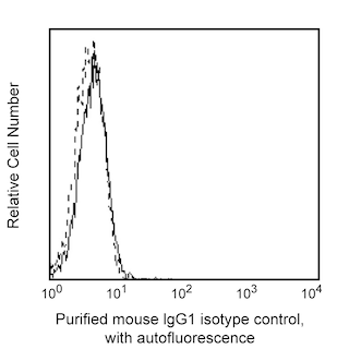
Clone SP34-2 is routinely tested using Purified Mouse IgG1 κ , clone MOPC-21 (Cat. No. Cat. No 556648 ), as the isotype control. Alternate isotype controls specific for the lambda light chain, such as clones S1-68.1 (Cat No. 553452) and A111-3 (Cat. No. 553485), are not routinely tested for flow cytometry application. Investigators are encouraged to validate the alternative clones for the desired applications.
Product Notices
- Since applications vary, each investigator should titrate the reagent to obtain optimal results.
- An isotype control should be used at the same concentration as the antibody of interest.
- Caution: Sodium azide yields highly toxic hydrazoic acid under acidic conditions. Dilute azide compounds in running water before discarding to avoid accumulation of potentially explosive deposits in plumbing.
- Sodium azide is a reversible inhibitor of oxidative metabolism; therefore, antibody preparations containing this preservative agent must not be used in cell cultures nor injected into animals. Sodium azide may be removed by washing stained cells or plate-bound antibody or dialyzing soluble antibody in sodium azide-free buffer. Since endotoxin may also affect the results of functional studies, we recommend the NA/LE (No Azide/Low Endotoxin) antibody format, if available, for in vitro and in vivo use.
- Species cross-reactivity detected in product development may not have been confirmed on every format and/or application.
- Please refer to www.bdbiosciences.com/us/s/resources for technical protocols.
Companion Products

.png?imwidth=320)
Clone SP34-2 is a mouse IgG1 isotype monoclonal antibody, descendant of SP34 (mouse IgG3), with the same specificity and reactivity pattern as the parent clone. It cross-reacts with a major subset of peripheral blood lymphocytes, but not monocytes or granulocytes, of baboon, and rhesus, cynomolgus, and pigtail macaque monkeys. The distribution on lymphocytes is similar to that observed with normal human donor lymphocytes with the majority of CD3-positive cells being negative when dual stained with antibodies to B or NK cells markers. SP34-2 is also capable of inducing cell proliferation on both human and non-human primate PBMC.
Development References (6)
-
Blumberg RS, Ley S, Sancho J, et al. Structure of the T-cell antigen receptor: evidence for two CD3 epsilon subunits in the T-cell receptor-CD3 complex. Proc Natl Acad Sci U S A. 1990; 87(18):7220-7224. (Clone-specific). View Reference
-
Carter DL, Shieh TM, Blosser RL et al. CD56 identifies monocytes and not natural killer cells in rhesus macaques. Cytometry. 1999; 37(1):41-50. (Biology). View Reference
-
Pessano S, Oettgen H, Bhan AK, Terhorst C. The T3/T cell receptor complex: antigenic distinction between the two 20-kd T3 (T3-delta and T3-epsilon) subunits. EMBO J. 1985; 4(2):337-344. (Immunogen). View Reference
-
Sancho J, Ledbetter JA, Choi MS, Kanner SB, Deans JP, Terhorst C. CD3-zeta surface expression is required for CD4-p56lck-mediated upregulation of T cell antigen receptor-CD3 signaling in T cells. J Biol Chem. 1992; 267(11):7871-7879. (Biology). View Reference
-
Schlossman SF. Stuart F. Schlossman .. et al., ed. Leucocyte typing V : white cell differentiation antigens : proceedings of the fifth international workshop and conference held in Boston, USA, 3-7 November, 1993. Oxford: Oxford University Press; 1995.
-
Wilson AD, Shooshtari M, Finerty S, Watkins P, Morgan AJ. Selection of monoclonal antibodies for the identification of lymphocyte surface antigens in the New World primate Saguinus oedipus oedipus (cotton top tamarin). J Immunol Methods. 1995; 178(2):195-200. (Biology). View Reference
Please refer to Support Documents for Quality Certificates
Global - Refer to manufacturer's instructions for use and related User Manuals and Technical data sheets before using this products as described
Comparisons, where applicable, are made against older BD Technology, manual methods or are general performance claims. Comparisons are not made against non-BD technologies, unless otherwise noted.
For Research Use Only. Not for use in diagnostic or therapeutic procedures.