Preparation And Storage
Recommended Assay Procedures
The PE conjugated D7715A7 antibody can be used for the immunofluorescent staining and flow cytometric analyses of mouse peripheral blood leukocytes and cell lines which express IL-6 receptors.
1. To help block nonspecific staining due to Fc receptors, preincubate ~1 million cells with 1 µg of the purified anti-CD32/CD16 (anti-FcγII/III receptor) antibody (clone 2.4G2, Fc Block™, Cat. No. 553142/553143) for 30 minutes at 4°C.
2. Incubate the cells with 0.12 - 2.0 µg of PE conjugated D7715A7 antibody (Cat. No. 554462) at 4°C for 45 minutes. Wash cells three times with staining medium containing sodium azide (e.g., Dulbecco's PBS or tissue culture medium [without phenol red] with 0.09% sodium azide and 1% heat-inactivated FCS). We encourage investigators to titrate the D7715A7 antibody up to saturating levels for optimal performance, minimizing the risk for dim staining.
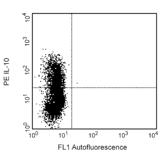
3. Resuspend cells in staining medium and analyze by flow cytometry using appropriate specificity and compensation controls. Using this method, positive staining was seen with the B9 cell line but not with the mouse MC/9 mast cell line and C20.4 CD4+ T cell line.
Product Notices
- Since applications vary, each investigator should titrate the reagent to obtain optimal results.
- Please refer to www.bdbiosciences.com/us/s/resources for technical protocols.
- Caution: Sodium azide yields highly toxic hydrazoic acid under acidic conditions. Dilute azide compounds in running water before discarding to avoid accumulation of potentially explosive deposits in plumbing.
- For fluorochrome spectra and suitable instrument settings, please refer to our Multicolor Flow Cytometry web page at www.bdbiosciences.com/colors.
- An isotype control should be used at the same concentration as the antibody of interest.
Companion Products

The D7715A7 monoclonal antibody specifically binds to the mouse IL-6 receptor, which is also known as, CD126. The immunogen used to generate the D7715A7 hybridoma was OKT4 hybridoma cells. The D7715A7 hybridoma was selected based on its capacity to produce antibody that inhibited IL-6 binding to 2F4 cells (mouse B-cell hybridoma expressing high levels of IL-6 receptors). The binding of purified D7715A7 to B9 cells (mouse B-cell hybridoma expressing high levels of IL-6 receptors) is inhibited by recombinant mouse IL-6. This antibody has been reported to inhibit the in vitro and in vivo growth of the IL-6-dependent plasmacytoma line, T1033C2.

Development References (3)
-
Coulie PG, Vink A, Van Snick J. A monoclonal antibody specific for the murine IL-6-receptor inhibits the growth of a mouse plasmacytoma in vivo. Curr Top Microbiol Immunol. 1990; 166:43-46. (Clone-specific). View Reference
-
Vink A, Coulie P, Warnier G, et al. Mouse plasmacytoma growth in vivo: enhancement by interleukin 6 (IL-6) and inhibition by antibodies directed against IL-6 or its receptor. J Exp Med. 1990; 172(3):997-1000. (Biology). View Reference
-
Zola H. Detection of receptors for cytokines and growth factors. The Immunologist. 1994:47.
Please refer to Support Documents for Quality Certificates
Global - Refer to manufacturer's instructions for use and related User Manuals and Technical data sheets before using this products as described
Comparisons, where applicable, are made against older BD Technology, manual methods or are general performance claims. Comparisons are not made against non-BD technologies, unless otherwise noted.
For Research Use Only. Not for use in diagnostic or therapeutic procedures.