The site is undergoing maintenance on
Thursday, September 10, 6:00 pm through Friday, September 11, 2:00 am (CEST), 2026.
If you encounter problems while ordering, please contact us at BDBCustomerService@bd.com
See More
The site is undergoing maintenance on
Thursday, September 10, 6:00 pm through Friday, September 11, 2:00 am (CEST), 2026.
If you encounter problems while ordering, please contact us at BDBCustomerService@bd.com
See MoreThe site is undergoing maintenance on
Thursday, September 10, 6:00 pm through Friday, September 11, 2:00 am (CEST), 2026
.If you encounter problems while ordering, please contact us at BDBCustomerService@bd.com
This page has been recently translated and is available in French now.
Looks like you're visiting us from {countryName}.
Would you like to stay on the current location site or be switched to your location?
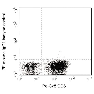
Flow cytometric analysis of CX3CL1 expression on wild type (Left Panel) or Crispr/Cas9 knockout (Right Panel) HepG2 cells. Wild type or knockout human HepG2 cells were detached with Accutase (Cat. No. 561527) and stained with either Mouse Anti-Human CX3CL1 antibody (Cat. No. 566905; solid line histogram) or PE mIgG1, κ Isotype Control (Cat. No. 556650; dashed line histogram). Fluorescence histograms were derived from gated events with the forward and side light-scatter characteristics of live cells. Flow cytometric analysis was performed using a BD™ Canto II Flow Cytometer System. Crispr/Cas9 knockout HepG2 was generated by Synthego Corp. Knockout HepG2 cells were bulk sorted and sorted cells were expanded by culturing before staining. Data shown on this Technical Data Sheet are not lot specific.
Any use of products other than the permitted use without the express written authorization of Becton, Dickinson and Company is strictly prohibited.
BD™ CompBeads can be used as surrogates to assess fluorescence spillover (Compensation). When fluorochrome conjugated antibodies are bound to CompBeads, they have spectral properties very similar to cells. However, for some fluorochromes there can be small differences in spectral emissions compared to cells, resulting in spillover values that differ when compared to biological controls. It is strongly recommended that when using a reagent for the first time, users compare the spillover on cell and CompBead to ensure that BD Comp beads are appropriate for your specific cellular application.
The V13-864 monoclonal antibody specifically recognizes CX3C motif chemokine Ligand 1 (CX3CL1), which is also known as fractalkine. CX3CL1 (Fractalkine) is the only known member of the CX3C chemokine subfamily and is expressed as both membrane-bound and secreted forms. Unlike other chemokines, CX3CL1is expressed on the surface of endothelial cells and not significantly expressed on peripheral blood leukocytes. CX3CL1's receptor is known as CX3CR1 and is expressed on cytotoxic T cells, NK cells, dendritic cells, and monocytes. Overexpression of CX3CL1 (Fractalkine) in some malignant tumors may contribute to the recruitment of effectors for innate and adaptive immunity.
Please refer to Support Documents for Quality Certificates
Global - Refer to manufacturer's instructions for use and related User Manuals and Technical data sheets before using this products as described
Comparisons, where applicable, are made against older BD Technology, manual methods or are general performance claims. Comparisons are not made against non-BD technologies, unless otherwise noted.
Please refer to Support Documents for Quality Certificates
Global - Refer to manufacturer's instructions for use and related User Manuals and Technical data sheets before using this products as described
Comparisons, where applicable, are made against older BD Technology, manual methods or are general performance claims. Comparisons are not made against non-BD technologies, unless otherwise noted.
For Research Use Only. Not for use in diagnostic or therapeutic procedures.