-
Your selected country is
Sweden
- Change country/language
Old Browser
This page has been recently translated and is available in French now.
Looks like you're visiting us from {countryName}.
Would you like to stay on the current country site or be switched to your country?
Multicolor flow cytometric analysis of CD49b (Integrin α2) expression on viable Mouse NK cells. C57BL/6 Mouse splenic leucocytes were preincubated with Purified Rat Anti-Mouse CD16/CD32 antibody (Mouse BD Fc Block™) [Cat. No. 553141/553142]. The cells were then stained with APC Mouse Anti-Mouse NK-1.1 antibody (Cat. No. 550627) and with either BD Horizon™ BV605 Hamster IgG1, κ Isotype Control (Cat. No. 563009; Left Plot) or BD Horizon™ BV605 Hamster Anti-Mouse CD49b (Integrin α2) [Cat. No. 569508; Right Plot] at 0.5 µg/test. DAPI (4',6-Diamidino-2-Phenylindole, Dihydrochloride) Solution (Cat. No. 564907) was added to cells right before analysis. The bivariate pseudocolor density plot showing the correlated expression of CD49b (Integrin α2) [or Ig Isotype control staining] versus NK1.1 was derived from gated events with the side and forward light-scatter characteristics of viable (DAPI-negative) leucocytes. Flow cytometry and data analysis were performed using a BD LSRFortessa™ X-20 Cell Analyzer System and FlowJo™ software. Data shown on this Technical Data Sheet are not lot specific.
BD Horizon™ BV605 Hamster Anti-Mouse CD49b (Integrin α2)
Regulatory Status Legend
Any use of products other than the permitted use without the express written authorization of Becton, Dickinson and Company is strictly prohibited.
Preparation And Storage
Recommended Assay Procedures
BD® CompBeads can be used as surrogates to assess fluorescence spillover (compensation). When fluorochrome conjugated antibodies are bound to BD® CompBeads, they have spectral properties very similar to cells. However, for some fluorochromes there can be small differences in spectral emissions compared to cells, resulting in spillover values that differ when compared to biological controls. It is strongly recommended that when using a reagent for the first time, users compare the spillover on cells and BD® CompBeads to ensure that BD® CompBeads are appropriate for your specific cellular application.
For optimal and reproducible results, BD Horizon Brilliant Stain Buffer should be used anytime BD Horizon Brilliant dyes are used in a multicolor flow cytometry panel. Fluorescent dye interactions may cause staining artifacts which may affect data interpretation. The BD Horizon Brilliant Stain Buffer was designed to minimize these interactions. When BD Horizon Brilliant Stain Buffer is used in in the multicolor panel, it should also be used in the corresponding compensation controls for all dyes to achieve the most accurate compensation. For the most accurate compensation, compensation controls created with either cells or beads should be exposed to BD Horizon Brilliant Stain Buffer for the same length of time as the corresponding multicolor panel. More information can be found in the Technical Data Sheet of the BD Horizon Brilliant Stain Buffer (Cat. No. 563794/566349) or the BD Horizon Brilliant Stain Buffer Plus (Cat. No. 566385).
Product Notices
- Please refer to www.bdbiosciences.com/us/s/resources for technical protocols.
- Please observe the following precautions: Absorption of visible light can significantly alter the energy transfer occurring in any tandem fluorochrome conjugate; therefore, we recommend that special precautions be taken (such as wrapping vials, tubes, or racks in aluminum foil) to prevent exposure of conjugated reagents, including cells stained with those reagents, to room illumination.
- Caution: Sodium azide yields highly toxic hydrazoic acid under acidic conditions. Dilute azide compounds in running water before discarding to avoid accumulation of potentially explosive deposits in plumbing.
- For fluorochrome spectra and suitable instrument settings, please refer to our Multicolor Flow Cytometry web page at www.bdbiosciences.com/colors.
- CF™ is a trademark of Biotium, Inc.
- Although every effort is made to minimize the lot-to-lot variation in the efficiency of the fluorochrome energy transfer, differences in the residual emission from BD Horizon™ BV421 may be observed. Therefore, we recommend that individual compensation controls be performed for every BD Horizon™ BV605 conjugate.
- BD Horizon Brilliant Violet 605 is covered by one or more of the following US patents: 8,110,673; 8,158,444; 8,227,187; 8,455,613; 8,575,303; 8,354,239.
- BD Horizon Brilliant Stain Buffer is covered by one or more of the following US patents: 8,110,673; 8,158,444; 8,575,303; 8,354,239.
- Please refer to http://regdocs.bd.com to access safety data sheets (SDS).
- Since applications vary, each investigator should titrate the reagent to obtain optimal results.
- An isotype control should be used at the same concentration as the antibody of interest.
Companion Products
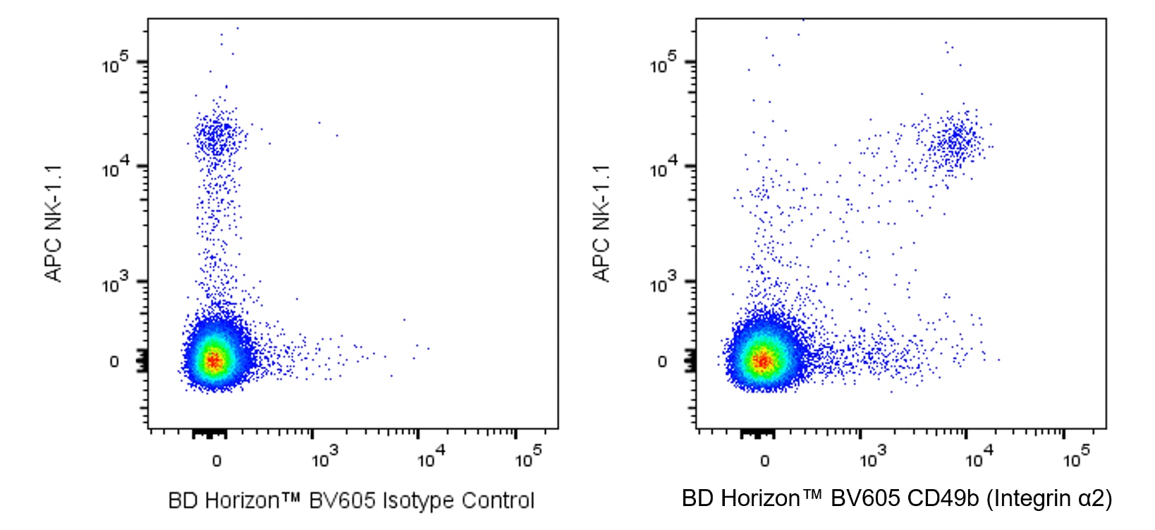
The HMα2 antibody reacts with integrin α2 chain (CD49b), the 150-kDa transmembrane glycoprotein that non-covalently associates with the integrin β1 subunit (CD29) to form the integrin α2β1 complex known as VLA-2. VLA-2, a receptor for collagen and laminin, is expressed on some splenic CD4+ T lymphocytes and NK-T cells, intestinal intraepithelial and lamina propria lymphocytes, splenic NK cells, epithelial cells, and platelets; but it is not on thymocytes or Peyer's-patch or lymphnode lymphocytes. The expression of VLA-2 is upregulated on lymphocytes in response to mitogens. The HMα2 antibody has been reported to partially block the interaction of T-cell blasts, but not NK cells, with collagen. Purified HMα2 mAb blocks the staining of splenic NK cells by the anti-CD49b/Pan-NK Cells mAb DX5 (Cat. No. 553858, for the PE conjugate). Therefore, mAb HMα2 may be used like the DX5 mAb for identification of NK cells.
Development References (5)
-
Arase H, Saito T, Phillips JH, Lanier LL. Cutting edge: the mouse NK cell-associated antigen recognized by DX5 monoclonal antibody is CD49b (alpha 2 integrin, very late antigen-2). J Immunol. 2001; 167(3):1141-1144. (Biology). View Reference
-
Chen H, Paul WE. A population of CD62Llow Nk1.1- CD4+ T cells that resembles NK1.1+ CD4+ T cells. Eur J Immunol. 1998; 28(10):3172-3182. (Clone-specific: Flow cytometry). View Reference
-
Miyake S, Sakurai T, Okumura K, Yagita H. Identification of collagen and laminin receptor integrins on murine T lymphocytes. Eur J Immunol. 1994; 24(9):2000-2005. (Immunogen: Blocking, Flow cytometry, Immunoprecipitation). View Reference
-
Noto K, Kato K, Okumura K, Yagita H. Identification and functional characterization of mouse CD29 with a mAb. Int Immunol. 1995; 7(5):835-842. (Biology). View Reference
-
Tanaka T, Ohtsuka Y, Yagita H, Shiratori Y, Omata M, Okumura K. Involvement of alpha 1 and alpha 4 integrins in gut mucosal injury of graft-versus-host disease. Int Immunol. 1995; 7(8):1183-1189. (Clone-specific: Flow cytometry). View Reference
Please refer to Support Documents for Quality Certificates
Global - Refer to manufacturer's instructions for use and related User Manuals and Technical data sheets before using this products as described
Comparisons, where applicable, are made against older BD Technology, manual methods or are general performance claims. Comparisons are not made against non-BD technologies, unless otherwise noted.
For Research Use Only. Not for use in diagnostic or therapeutic procedures.
Report a Site Issue
This form is intended to help us improve our website experience. For other support, please visit our Contact Us page.