Old Browser
This page has been recently translated and is available in French now.
Looks like you're visiting us from {countryName}.
Would you like to stay on the current country site or be switched to your country?


.png)

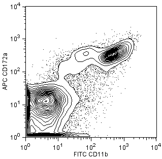
Flow cytometric analysis of APC-conjugated anti-mouse CD172a on mouse bone marrow. Bone marrow cells from BALB/c mice were stained with APC anti-mouse CD172a (clone P84, Cat. No. 560106) and FITC anti-mouse CD11b (Clone M1/70, Cat. No. 553310) and analyzed by flow cytometry. Flow cytometry was performed on a BD FACSCalibur™ System and the contour plot was derived from the gated events based on light scattering characteristics of viable bone marrow cells.
.png)

BD Pharmingen™ APC Rat anti-Mouse CD172a
.png)
Regulatory Status Legend
Any use of products other than the permitted use without the express written authorization of Becton, Dickinson and Company is strictly prohibited.
Preparation And Storage
Product Notices
- Since applications vary, each investigator should titrate the reagent to obtain optimal results.
- Caution: Sodium azide yields highly toxic hydrazoic acid under acidic conditions. Dilute azide compounds in running water before discarding to avoid accumulation of potentially explosive deposits in plumbing.
- For fluorochrome spectra and suitable instrument settings, please refer to our Multicolor Flow Cytometry web page at www.bdbiosciences.com/colors.
- Please refer to www.bdbiosciences.com/us/s/resources for technical protocols.
Companion Products
.png?imwidth=320)

The P84 monoclonal antibody specifically binds to CD172a, also known as SIgnal-Regulatory Protein α (SIRPα), Src Homology 2 domain-containing protein tyrosine Phosphatase (SHP) Substrate 1 (SHPS-1), or Brain Immunoglobulin-like molecule with Tyrosine-based activation motifs (BIT). CD172a is an adhesion molecule of the Ig superfamily which is expressed on neurons in the central nervous system and the retina, on macrophages, and on bone-marrow myeloid cells. Its ligand, CD47, or Integrin-Associated Protein (IAP), is expressed by a wide variety of cells. CD172a and CD47 are proposed to mediate bi-directional signaling to modify neural synaptic activity and regulate the phagocytic activities of macrophages.

Development References (6)
-
Chuang W, Lagenaur CF. Central nervous system antigen P84 can serve as a substrate for neurite outgrowth. Dev Biol. 1990; 137(2):219-232. (Immunogen). View Reference
-
Comu S, Weng W, Olinsky S. The murine P84 neural adhesion molecule is SHPS-1, a member of the phosphatase-binding protein family. J Neurosci. 1997; 17(22):8702-8710. (Biology). View Reference
-
Gresham HD, Dale BM, Potter JW, et al. Negative regulation of phagocytosis in murine macrophages by the Src kinase family member, Fgr. J Exp Med. 2000; 191(3):515-528. (Biology). View Reference
-
Mi ZP, Jiang P, Weng WL, Lindberg FP, Narayanan V, Lagenaur CF. Expression of a synapse-associated membrane protein, P84/SHPS-1, and its ligand, IAP/CD47, in mouse retina. J Comp Neurol. 2000; 416(3):335-344. (Biology). View Reference
-
Oldenborg PA, Zheleznyak A, Fang YF, Lagenaur CF, Gresham HD, Lindberg FP. Role of CD47 as a marker of self on red blood cells. Science. 2000; 288(5473):2051-2054. (Biology). View Reference
-
Veillette A, Thibaudeau E, Latour S. High expression of inhibitory receptor SHPS-1 and its association with protein-tyrosine phosphatase SHP-1 in macrophages. J Biol Chem. 1998; 273(35):22719-22728. (Biology). View Reference
Please refer to Support Documents for Quality Certificates
Global - Refer to manufacturer's instructions for use and related User Manuals and Technical data sheets before using this products as described
Comparisons, where applicable, are made against older BD Technology, manual methods or are general performance claims. Comparisons are not made against non-BD technologies, unless otherwise noted.
For Research Use Only. Not for use in diagnostic or therapeutic procedures.
Report a Site Issue
This form is intended to help us improve our website experience. For other support, please visit our Contact Us page.