Old Browser
This page has been recently translated and is available in French now.
Looks like you're visiting us from {countryName}.
Would you like to stay on the current country site or be switched to your country?


.png)

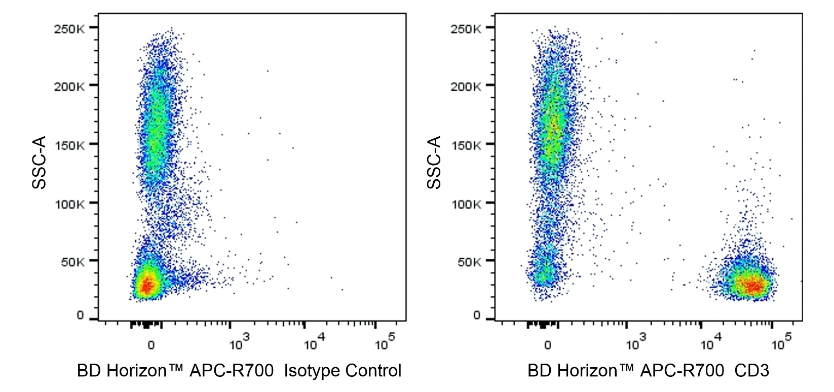
Multiparameter flow cytometric analysis of CD3 expression on human peripheral blood leucocytes. Human whole blood was stained with either BD Horizon™ APC-R700 Mouse IgG2a, κ Isotype Control (Cat. No. 565126; Left Plot) or BD Horizon APC-R700 Mouse Anti-Human CD3 antibody (Cat. No. 566796/566797; Right Plot). Erythrocytes were lysed with BD Pharm Lyse™ Lysing Buffer (Cat. No. 555899). Two-parameter pseudocolor density plots showing the correlated expression of CD3 (or Ig Isotype control staining) versus side light-scatter (SSC-A) signals were derived from gated events with the forward and side light-scatter characteristics of viable leucocyte populations. Flow cytometry and data analysis were performed using a BD LSRFortessa™ X-20 Cell Analyzer System and FlowJo™ software.
.png)

BD Horizon™ APC-R700 Mouse Anti-Human CD3
.png)
Regulatory Status Legend
Any use of products other than the permitted use without the express written authorization of Becton, Dickinson and Company is strictly prohibited.
Preparation And Storage
Product Notices
- This reagent has been pre-diluted for use at the recommended Volume per Test. We typically use 1 × 10^6 cells in a 100-µl experimental sample (a test).
- An isotype control should be used at the same concentration as the antibody of interest.
- Source of all serum proteins is from USDA inspected abattoirs located in the United States.
- Caution: Sodium azide yields highly toxic hydrazoic acid under acidic conditions. Dilute azide compounds in running water before discarding to avoid accumulation of potentially explosive deposits in plumbing.
- Alexa Fluor® is a registered trademark of Molecular Probes, Inc., Eugene, OR.
- For fluorochrome spectra and suitable instrument settings, please refer to our Multicolor Flow Cytometry web page at www.bdbiosciences.com/colors.
- Please refer to http://regdocs.bd.com to access safety data sheets (SDS).
- Please refer to www.bdbiosciences.com/us/s/resources for technical protocols.
Companion Products


.png?imwidth=320)
The OKT3 monoclonal antibody specifically recognizes the CD3 epsilon subunit (CD3e/CD3ε) of the CD3 complex which consists of four transmembrane proteins (γ, δ, ε, ζ) that are associated with the T cell antigen receptor (TCR) to form the CD3/TCR complex. The CD3 complex associates with either TCR αβ or TCR γδ heterodimers that are alternatively expressed by some thymocytes, T cells or NKT cells. The CD3 complex is required for the cell surface expression and signal-transducing functions of the TCR. The CD3 complex is expressed by ~60-85% thymocytes and by all peripheral mature T cells. CD3e is also known as T3E or TCRE. CD3e is a ~20 kDa unglycosylated type I transmembrane protein that is encoded by CD3E which belongs to the immunoglobulin superfamily (IgSF). CD3e has an Ig-like extracellular domain (ECD) and an immunoreceptor tyrosine-based activation motif (ITAM) in its cytoplasmic domain. The OKT3 antibody can reportedly fix complement, stimulate T cell proliferation and cytokine production, and block the binding of other human CD3e-specific antibodies including UCHT1 and SK7.
This antibody was conjugated to BD Horizon APC-R700, which has been developed exclusively by BD Biosciences as a better alternative to Alexa Fluor® 700. APC-R700 excites and emits at similar wavelengths to Alexa Fluor® 700 yet exhibits significantly improved brightness. This dye can be excited by the red laser and detected with the same filter set as Alexa Fluor® (eg, 730/45-nm filter).

Development References (12)
-
Burns GF, Boyd AW, Beverley PC. Two monoclonal anti-human T lymphocyte antibodies have similar biologic effects and recognize the same cell surface antigen. J Immunol. 1982; 129(4):1451-1457. (Clone-specific: Blocking, Functional assay, Immunoprecipitation, Radioimmunoassay). View Reference
-
Emmrich F. Selective stimulation of human CD4 and CD8 T-cells by crosslining the T-cell receptor with subset-specific differentiation antigens. In: McMichael AJ. A.J. McMichael .. et al., ed. Leucocyte typing III : white cell differentiation antigens. Oxford New York: Oxford University Press; 1987:203-206.
-
Ernst DN, Shih CC. CD3 complex. J Biol Regul Homeost Agents. 2000; 14(3):226-229. (Biology). View Reference
-
Horibe K, Knowles RW, Naito K, Morishima Y, Dupont B. Analysis of T lymphocyte antibody specificities: Comparison of serology with immunoprecipitation patterns. In: Bernard A. A. Bernard .. et al., ed. Leucocyte typing : human leucocyte differentiation antigens detected by monoclonal antibodies. Berlin New York: Springer-Verlag; 1984:212-224.
-
Kung P, Goldstein G, Reinherz EL, Schlossman SF. Monoclonal antibodies defining distinctive human T cell surface antigens. Science. 1979; 206(4416):347-349. (Immunogen: Cytotoxicity, Flow cytometry, Radioimmunoassay). View Reference
-
Kurrle R, Seyfert W, Trautwein A, Seiler FR. T cell activation by CD3 antibodies. In: Reinherz EL. Ellis L. Reinherz .. et al., ed. Leukocyte typing II. New York: Springer-Verlag; 1986:137-146.
-
Li B, Wang H, Dai J, et al. Construction and characterization of a humanized anti-human CD3 monoclonal antibody 12F6 with effective immunoregulation functions. Immunology. 2005; 116(4):487-498. (Clone-specific: Blocking, Flow cytometry). View Reference
-
Semnani R, Nutman TB, Corrado G, Hochman P, Shaw S, Van Seventer GA. Costimulation mediated by purified ICAM-1 and LFA-3 regulates differential stimulation and cytokine secretion of human 'naive' and 'memory' CD4+ T cells. In: Schlossman SF. Stuart F. Schlossman .. et al., ed. Leucocyte typing V : white cell differentiation antigens. Oxford: Oxford University Press; 1995:1488-1491.
-
Touraine JL, Favrot MC, Ansary ME, Cordier G, de bouteiller O. Phenotype of prothymocytes from human bone marrow determined by monoclonal antibodies: Modification induced by thymic factots. In: Bernard A. A. Bernard .. et al., ed. Leucocyte typing : human leucocyte differentiation antigens detected by monoclonal antibodies. Berlin New York: Springer-Verlag; 1984:298-311.
-
Tunnacliffe A, Olsson C, Traunecker A, Krissansen GW, Karjalainen K, de la Hera A. The majority of CD3 epitopes are conferred by the epsilon chain. In: Knapp W. W. Knapp .. et al., ed. Leucocyte typing IV : white cell differentiation antigens. Oxford New York: Oxford University Press; 1989:295-296.
-
Van Wauwe JP, De Mey JR, Goossens JG. OKT3: a monoclonal anti-human T lymphocyte antibody with potent mitogenic properties. J Immunol. 1980; 124:2708-2713. (Clone-specific: Functional assay). View Reference
-
Van Wauwe JP, Goossens JG, Beverley PC. Human T lymphocyte activation by monoclonal antibodies; OKT3, but not UCHT1, triggers mitogenesis via an interleukin 2-dependent mechanism. J Immunol. 1984; 133(1):129-132. (Clone-specific: Functional assay). View Reference
Please refer to Support Documents for Quality Certificates
Global - Refer to manufacturer's instructions for use and related User Manuals and Technical data sheets before using this products as described
Comparisons, where applicable, are made against older BD Technology, manual methods or are general performance claims. Comparisons are not made against non-BD technologies, unless otherwise noted.
For Research Use Only. Not for use in diagnostic or therapeutic procedures.
Report a Site Issue
This form is intended to help us improve our website experience. For other support, please visit our Contact Us page.