Old Browser
This page has been recently translated and is available in French now.
Looks like you're visiting us from {countryName}.
Would you like to stay on the current country site or be switched to your country?
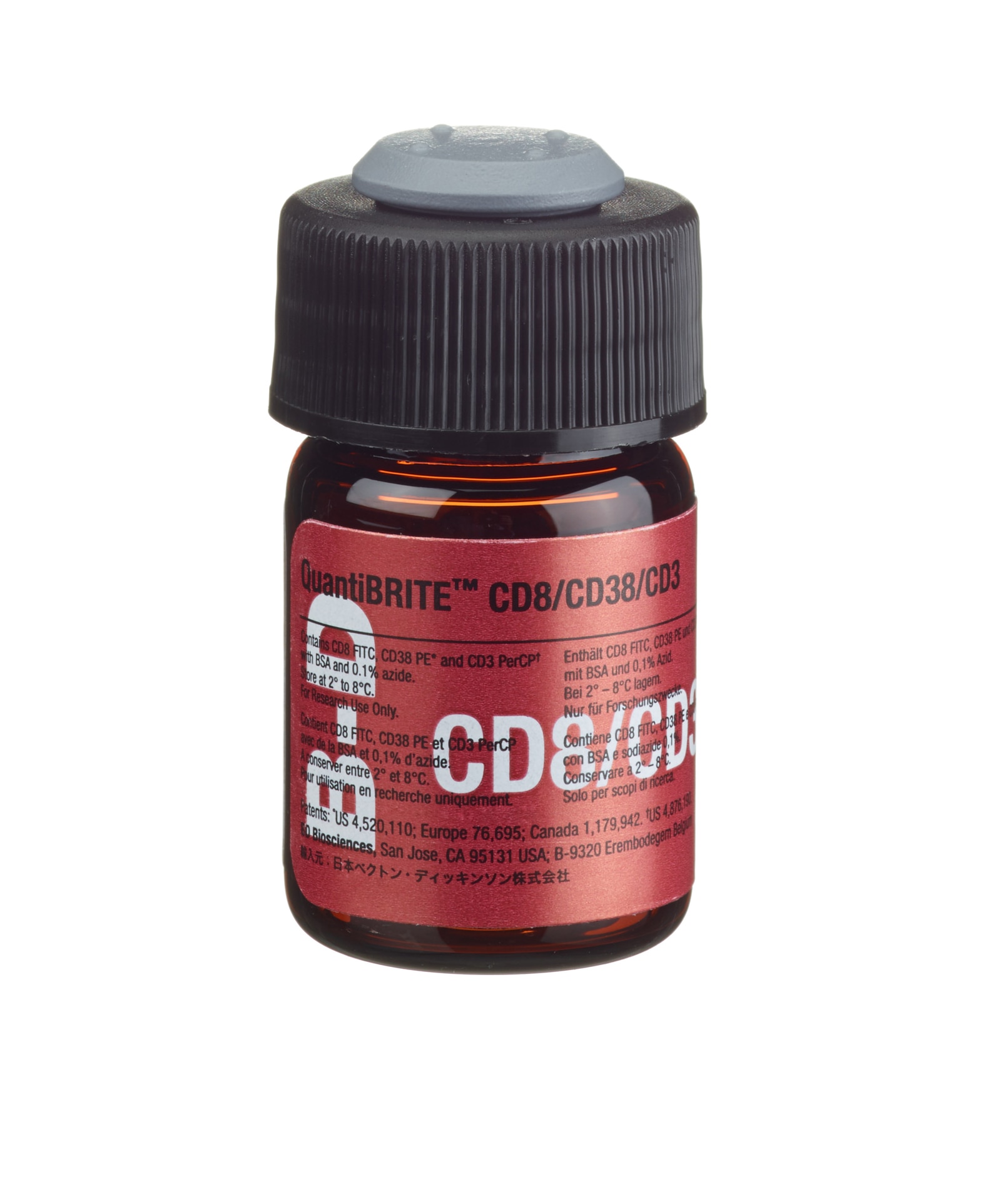
BD Quantibrite™ Anti-Human CD8 FITC/CD38 PE/CD3 PerCP
(RUO (GMP))

Anti-Human CD8 FITC/CD38 PE/CD3 PerCP
Regulatory Status Legend
Any use of products other than the permitted use without the express written authorization of Becton, Dickinson and Company is strictly prohibited.
Description
CD8, clone SK1, is derived from hybridization of mouse NS-1 myeloma cells with spleen cells from BALB/c mice immunized with human peripheral blood T lymphocytes. CD38, clone HB7, is derived from hybridization of mouse P3-X63-Ag8.653 myeloma cells with spleen cells from BALB/c mice immunized with the BJAB cell line. CD3, clone SK7, is derived from hybridization of mouse NS-1 myeloma cells with spleen cells from BALB/c mice immunized with human thymocytes. CD8 recognizes an antigen expressed on the 32-kilodalton (kd) α subunit of a disulfide-linked bimolecular complex. The cytoplasmic domain of the α subunit of the CD8 antigen is associated with the protein tyrosine kinase p56lck. The CD8 molecule interacts with class I major histocompatibility complex (MHC) molecules resulting in increased adhesion between the CD8+ T lymphocytes and the target cells. Binding of the CD8 molecule to class I MHC molecules enhances the activation of resting T lymphocytes. CD38 recognizes an integral membrane glycoprotein, Mr 45 kd, with a protein core of 35 kd. CD3 recognizes the epsilon chain of the CD3 antigen/T-cell antigen receptor (TCR) complex. This complex is composed of at least six proteins that range in molecular weight from 20–30 kd. The antigen recognized by the CD3 antibody is noncovalently associated with either α/β or γ/δ TCR (70–90 kd).
Preparation And Storage
The QuantiBRITE CD8/CD38/CD3 reagent is supplied as a combination of CD8 FITC, CD38 PE (≥95% 1:1 PE:mAb ratio), and CD3 PerCP in 1 mL of PBS containing bovine serum albumin and 0.1% sodium azide. Vials should be stored at 2–8°C. QuantiBRITE reagents should not be frozen and should be protected from prolonged exposure to light. The reagent is stable for the period shown on the bottle label when stored as directed.
| Description | Clone | Isotype | EntrezGene ID |
|---|---|---|---|
| CD8 FITC | SK1 | IgG1, κ | N/A |
| CD38 PE | HB7 | IgG1, κ | N/A |
| CD3 PerCP | SK7 | IgG1, κ | N/A |
Development References (44)
-
Babusikova O, Ondrackova V, Prachar J, Kusenda J, Hraska V. Flow cytometric analysis of some activation/proliferation markers on human thymocytes and their correlation with cell proliferation. Neoplasma. 1999; 46(6):349-355. (Biology).
-
Badley AD, Parato K, Cameron DW, et al. Dynamic correlation of apoptosis and immune activation during treatment of HIV infection. Cell Death Differ. 1999; 6(5):420-432. (Biology).
-
Benito J, Zabay J, Gil J, et al. Quantitative alterations of the functionally distinct subsets of CD4 and CD8 T lymphocytes in asymptomatic HIV infection: Changes in the expression of CD45RO, CD45RA, CD11b, CD38, HLA-DR, and CD35 antigens. J Acquir Immune Defic Syndr Hum Retrovirol. 1997; 14:128-135. (Biology).
-
Brenner M, Groh V, Porcelli A, et al. Knapp W, Dörken B, Gilks W, et al, ed. Leucocyte Typing IV: White Cell Differentiation Antigens. 1989:1049-1053.
-
Clevers H, Alarcón B, Wileman T, Terhorst C. The T cell receptor/CD3 complex: a dynamic protein ensemble. Annual Rev Immunol. 1988; 6:629. (Biology).
-
Damle RN, Wasil T, Fais F, et al. Ig V gene mutation status and CD38 expression as novel prognostic indicators in chronic lymphocytic leukemia. Blood. 1999; 94(6):1840-1847. (Biology).
-
Dörken B, Möller P, Pezzutto A, Schwartz-Albiez R, et al. Knapp W, Dörken B, Gilks W, ed. Leucocyte Typing IV: White Cell Differentiation Antigens. New York: Oxford University Press; 1989:86.
-
Evans RL, Wall DW, Platsoucas CD, et al. Thymus-dependent membrane antigens in man: inhibition of cell-mediated lympholysis by monoclonal antibodies to TH2 antigen. Proc Natl Acad Sci U S A. 1981; 78(1):544-548. (Biology). View Reference
-
Gallagher PF, Fazekas de St. Groth B, Miller JFAP. CD4 and CD8 molecules can physically associate with the same T-cell receptor. Proc Natl Acad Sci USA. 1989; 86:10044-10048. (Biology).
-
Garson JA, Beverley PCL, Coakham HB, Harper EJ. Monoclonal antibodies against human T lymphocytes label Purkinje neurones of many species. Nature. 1982; 298:375-377. (Biology).
-
Giorgi J, Hultin L. Lymphocyte subset alterations and immunophenotyping by flow cytometry in HIV disease. Clin Immunol Newslett. 1990; 10(4):55-61. (Biology).
-
Giorgi JV, Detels R. T-cell subset alterations in HIV-infected homosexual men: NIAID multicenter AIDS cohort study. Clin Immunol Immunopathol. 1989; 52:10-18. (Biology).
-
Giorgi JV, Ho H-N, Hirji K, et al. CD8+ lymphocyte activation at human immunodeficiency virus type 1 seroconversion: development of HLA-DR+CD38-CD8+ cells is associated with subsequent stable CD4+ cell levels. The Multicenter AIDS Cohort Study Group. J Infect Dis. 1994; 170:775-781. (Biology).
-
Giorgi JV, Liu Z, Hultin LE, Cumberland WG, Hennessey K, Detels R. Elevated levels of CD38<up>+CD8+ T cells in HIV infection add to the prognostic value of low CD4+ T cell levels: results of 6 years of follow-up. J Acquir Immune Defic Syndr. 1993; 6:904-912. (Biology).
-
Haynes BF. Summary of T-cell studies performed during the Second International Workshop and Conference on Human Leukocyte Differentiation Antigens. In: Reinherz EL. Ellis L. Reinherz .. et al., ed. Leukocyte typing II. New York: Springer-Verlag; 1986:3-30.
-
Ho HN, Hultin LE, Mitsuyasu RT, et al. Circulating HIV-specific CD8+ cytotoxic T cells express CD38 and HLA-DR antigens. J Immunol. 1993; 150:3070-3079. (Biology).
-
Hultin LE, Matud JL, Giorgi JV. Quantitation of CD38 activation antigen expression on CD8+ T cells in HIV-1 infection using CD4 expression on CD4+ T lymphocytes as a biological calibrator. Cytometry. 1998; 33(2):123-132. (Biology).
-
Inoue K, Ogawa H, Sonoda Y, et al. Aberrant overexpression of the Wilms tumor gene (WT1) in human leukemia. Blood. 1997; 89(4):1405-1412. (Biology).
-
Iyer SB, Hultin LE, Zawadzki JA, Davis KA, Giorgi JV Quantitation of CD38 expression using QuantiBRITE beads. Cytometry. 1998; 33(2):206-212. (Biology).
-
Kawagoe H, Humphries RK, Blair A, Sutherland HJ, Hogge DE. Expression of HOX genes, HOX cofactors, and MLL in phenotypically and functionally defined subpopulations of leukemic and normal human hematopoietic cells. Leukemia. 1999; 13(5):687-698. (Biology).
-
Kestens L, Vanham G, Gigase P, et al. Expression of activation antigens, HLA-DR and CD38, on CD8 lymphocytes during HIV-1 infection. AIDS. 1992; 6:793-797. (Biology).
-
Knowles RW. Immunochemical analysis of the T-cell–specific antigens. In: Reinherz EL. Ellis L. Reinherz .. et al., ed. Leukocyte typing II. New York: Springer-Verlag; 1986:259-288.
-
Kotzin BL, Benike CJ, Engleman EG. Induction of immunoglobulin-secreting cells in the allogeneic mixed leukocyte reaction: regulation by helper and suppressor lymphocyte subsets in man. J Immunol. 1981; 127(9):931-935. (Biology). View Reference
-
Landay A, Ohlsson-Wilhelm B, Giorgi JV. Application of flow cytometry to the study of HIV infection. AIDS. 1990; 4(6):479-497. (Biology). View Reference
-
Lanier LL, Allison JP, Phillips JH. Correlation of cell surface antigen expression on human thymocytes by multi-color flow cytometric analysis: implications for differentiation. J Immunol. 1986; 137(8):2501-2507. (Biology). View Reference
-
Lanier LL, Le AM, Phillips JH, Warner NL, Babcock GF. Subpopulations of human natural killer cells defined by expression of the Leu-7 (HNK-1) and Leu-11 (NK-15) antigens. J Immunol. 1983; 131(4):1789-1796. (Biology). View Reference
-
Lenki R, Bratt G, Holmberg V, Muirhead K, Sandstrom E. Indicators of T-cell activation: Correlation between quantitative CD38 expression and soluble CD8 levels in asymptomatic HIV+ individuals and healthy controls. Cytometry. 1998; 33:115-122. (Biology).
-
Moebius U. Knapp W, Dörken B, Gilks W, et al, ed. Leucocyte Typing IV. White Cell Differentiation Antigens. New York: Oxford University Press; 1989:342-343.
-
Otawa M, Kawanishi Y, Iwase O, Shoji N, Miyazawa K, Ohyashiki K. Comparative multi-color flow cytometric analysis of cell surface antigens in bone marrow hematopoietic progenitors between refractory anemia and aplastic anemia. Leuk Res. 2000; 24(4):359-366. (Biology).
-
Perfetto SP, Malone JD, Hawkes C, et al. CD38 expression on cryopreserved CD8+ T cells predicts HIV disease progression. Cytometry. 1998; 33(2):133-137. (Biology).
-
Pezzutto A, Behm F, Callard RE. Flow cytometry analysis of the B-cell blind panel: joint report. In: Knapp W. W. Knapp .. et al., ed. Leucocyte typing IV : white cell differentiation antigens. Oxford New York: Oxford University Press; 1989:165-174.
-
Rawstron AC, Owen RG, Davies FE, et al. Circulating plasma cells in multiple myeloma: characterization and correlation with disease stage. Br J Haematol. 1997; 97:46-55. (Biology).
-
Reichert T, DeBruyere M, Deneys V, et al. Lymphocyte subset reference ranges in adult Caucasians. Clin Immunol Immunopathol. 1991; 60(2):190-208. (Biology). View Reference
-
Reinherz EL, Kung PC, Goldstein G, Levey RH, Schlossman SF. Discrete stages of human intrathymic differentiation: analysis of normal thymocytes and leukemic lymphoblasts of T-cell lineage. Proc Natl Acad Sci U S A. 1980; 77(3):1588-1592. (Biology). View Reference
-
Rudd CE, Burgess KE, Barber EK, Schlossman SF. Knapp W, Dörken B, Gilks WR, et al, ed. Leucocyte Typing IV: White Cell Differentiation Antigens. New York, NY: Oxford University Press; 1989:326-327.
-
Schmitz JL, Czerniewski MA, Edinger M, et al. Multisite comparison of methods for the quantitation of the surface expression of CD38 on CD8(+) T lymphocytes. Cytometry. 2000; 42(3):174-179. (Biology).
-
Shinjo K, Takeshita A, Higuchi M, Ohnishi K, Ohno R. Erythropoietin receptor expression on human bone marrow erythroid precursor cells by a newly-devised quantitative flow-cytometric assay. Br J Haematol. 1997; 96(3):551-558. (Biology).
-
Tedder TF, Clement LT, Cooper MD. Discontinuous expression of a membrane antigen (HB-7) during B lymphocyte differentiation. Tissue Antigens. 1984; 24(3):140-149. (Biology). View Reference
-
Tedder TF, Crain MJ, Kubagawa H, Clement LT, Cooper MD. Evaluation of lymphocyte differentiation in primary and secondary immunodeficiency diseases. J Immunol. 1985; 135(3):1785-1791. (Biology). View Reference
-
Terstappen LW, Hollander Z, Meiners H, Loken MR. Quantitative comparison of myeloid antigens on five lineages of mature peripheral blood cells. J Leukoc Biol. 1990; 48(2):138-148. (Biology). View Reference
-
Terstappen LW, Huang S, Picker LJ. Flow cytometric assessment of human T-cell differentiation in thymus and bone marrow. Blood. 1992; 79(3):666-677. (Biology). View Reference
-
Terstappen LW, Huang S, Safford M, Lansdorp PM, Loken MR. Sequential generations of hematopoietic colonies derived from single nonlineage-committed CD34+ CD38– progenitor cells. Blood. 1991; 77:1218-1227. (Biology).
-
Vigano A, Saresella M, Rusconi S, Ferrante P, Clerici M. Expression of CD38 on CD8 T cells predicts maintenance of high viraemia in HAART-treated HIV-1-infected children. The Lancet. 1998; 352:1906-1906. (Biology).
-
Wood GS, Warner NL, Warnke RA. Anti–Leu-3/T4 antibodies react with cells of monocyte/macrophage and Langerhans lineage. J Immunol. 1983; 131(1):212-216. (Biology). View Reference
Please refer to Support Documents for Quality Certificates
Global - Refer to manufacturer's instructions for use and related User Manuals and Technical data sheets before using this products as described
Comparisons, where applicable, are made against older BD Technology, manual methods or are general performance claims. Comparisons are not made against non-BD technologies, unless otherwise noted.
For Research Use Only. Not for use in diagnostic or therapeutic procedures.
Although not required, these products are manufactured in accordance with Good Manufacturing Practices.
Report a Site Issue
This form is intended to help us improve our website experience. For other support, please visit our Contact Us page.