-
Reagents
- Flow Cytometry Reagents
-
Western Blotting and Molecular Reagents
- Immunoassay Reagents
-
Single-Cell Multiomics Reagents
- BD® OMICS-Guard Sample Preservation Buffer
- BD® AbSeq Assay
- BD® Single-Cell Multiplexing Kit
- BD Rhapsody™ ATAC-Seq Assays
- BD Rhapsody™ Whole Transcriptome Analysis (WTA) Amplification Kit
- BD Rhapsody™ TCR/BCR Next Multiomic Assays
- BD Rhapsody™ Targeted mRNA Kits
- BD Rhapsody™ Accessory Kits
- BD® OMICS-One Protein Panels
-
Functional Assays
-
Microscopy and Imaging Reagents
-
Cell Preparation and Separation Reagents
-
- BD® OMICS-Guard Sample Preservation Buffer
- BD® AbSeq Assay
- BD® Single-Cell Multiplexing Kit
- BD Rhapsody™ ATAC-Seq Assays
- BD Rhapsody™ Whole Transcriptome Analysis (WTA) Amplification Kit
- BD Rhapsody™ TCR/BCR Next Multiomic Assays
- BD Rhapsody™ Targeted mRNA Kits
- BD Rhapsody™ Accessory Kits
- BD® OMICS-One Protein Panels
- Norway (English)
-
Change country/language
Old Browser
This page has been recently translated and is available in French now.
Looks like you're visiting us from United States.
Would you like to stay on the current country site or be switched to your country?
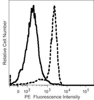
BD® AbSeq Oligo Mouse Anti-H2AX (pS139)
Clone N1-431 (RUO)


Regulatory Status Legend
Any use of products other than the permitted use without the express written authorization of Becton, Dickinson and Company is strictly prohibited.
Preparation And Storage
Recommended Assay Procedures
Put all BD® AbSeq reagents to be pooled into a Latch Rack for 500 µL Tubes (Thermo Fisher Scientific Cat. No. 4900). Arrange the tubes so that they can be easily uncapped and re-capped with an 8-Channel Screw Cap Tube Capper (Thermo Fisher Scientific Cat. No. 4105MAT) and the reagents aliquoted with a multi-channel pipette. BD® AbSeq tubes should be centrifuged for = 30 seconds at 400 × g to ensure removal of any content in the cap/tube threads prior to the first opening.
When using BD® AbSeq intracellular markers with the Single Cell 3' Sequencing Intracellular CITE-seq, cells must first be fixed and permeabilized using the BD Rhapsody™ Intracellular AbSeq Buffer Kit before the antibody-oligo can bind to the protein. Refer to the list of required companion products below and see BD Rhapsody™ System Single-Cell Labelling with BD® AbSeq Ab-Oligos for Intracellular CITE-seq (Doc ID: 23-24464) for the complete BD® AbSeq intracellular multiomics staining protocol. Contact your local Field Application Specialist (FAS) for additional guidance.
Use standard laboratory safety protocols. Read and understand the safety data sheets (SDSs) before handling chemicals. To obtain SDSs, go to regdocs.bd.com or contact BD Biosciences technical support at scomix@bdscomix.bd.com.
Warning: All biological specimens and materials contacting them are considered biohazardous. Handle as if capable of transmitting infection and dispose of with proper precautions in accordance with federal, state, and local regulations. Never pipette by mouth. Wear suitable protective clothing, eyewear, and gloves.
Product Notices
- Please refer to www.bdbiosciences.com/us/s/resources for technical protocols.
- This reagent has been pre-diluted for use at the recommended volume per test. Typical use is 2 µl for 1 × 10^6 cells in a 200-µl staining reaction.
- Caution: Sodium azide yields highly toxic hydrazoic acid under acidic conditions. Dilute azide compounds in running water before discarding to avoid accumulation of potentially explosive deposits in plumbing.
- The production process underwent stringent testing and validation to assure that it generates a high-quality conjugate with consistent performance and specific binding activity. However, verification testing has not been performed on all conjugate lots.
- Source of all serum proteins is from USDA inspected abattoirs located in the United States.
- Species cross-reactivity detected in product development may not have been confirmed on every format and/or application.
- Please refer to http://regdocs.bd.com to access safety data sheets (SDS).
- Please refer to bd.com/genomics-resources for technical protocols.
- Illumina is a trademark of Illumina, Inc.
- For U.S. patents that may apply, see bd.com/patents.
Data Sheets
Companion Products


Histones are highly basic proteins that complex with DNA to form chromatin. The H2AX histone (~15 kDa calculated molecular weight) is a member of the H2A histone family whose members are components of nucleosomal histone octamers. Double-stranded breaks in DNA caused by replication errors, apoptosis, or other physiological processes (including, immunoglobulin and TCR gene recombinations) and DNA damage caused by ionizing radiation, UV light, or cytotoxic agents lead to phosphorylation of H2AX on serine 139. H2AX (pS139) is also referred to as H2AX (pS140) when the N-terminal methionine that is normally excised during posttranslational processing is included in amino acid sequence numbering. Kinases such as ataxia telangiectasia mutated (ATM) or ATM-Rad3-related (ATR) phosphorylate H2AX to induce its function. Phosphorylated H2AX (also termed, gamma-H2AX) functions to recruit and localize DNA repair proteins or cell cycle checkpoint factors to the DNA-damaged sites. In this way, phosphorylated H2AX promotes DNA repair and maintains genomic stability and thus helps prevent oncogenic transformations. Immunofluorescent staining and bioimaging analysis of cultured cells can be used to readily identify H2AX (pS139)-containing foci. As such, H2AX (pS139) immunofluorescence localization serves as a biomarker for nuclear sites of DNA damage (e.g., double-stranded DNA breaks) in affected cells.
Development References (7)
-
Austin WR, Armijo AL, Campbell DO, et al.. Nucleoside salvage pathway kinases regulate hematopoiesis by linking nucleotide metabolism with replication stress. J Exp Med. 2012; 209(12):2215-2228. (Clone-specific: Flow cytometry). View Reference
-
Burma S, Chen BP, Murphy M, Kurimasa A, Chen DJ. ATM phosphorylates histone H2AX in response to DNA double-strand breaks. J Biol Chem. 2001; 276(45):42462-42467. (Biology). View Reference
-
Fernandez-Capetillo O, Lee A, Nussenzweig M, Nussenzweig A. H2AX: the histone guardian of the genome. DNA Repair (Amst). 2004; 3(8-9):959-967. (Biology). View Reference
-
Kuo LJ, Yang LX. Gamma-H2AX - A novel biomarker for DNA double-strand breaks. In Vivo. 2008; 22(3):305-309. (Biology). View Reference
-
Lavelle D, Vaitkus K, Ling Y, et al. Effects of tetrahydrouridine on pharmacokinetics and pharmacodynamics of oral decitabine. Blood. 2012; 119(5):1240-1247. (Clone-specific: Flow cytometry). View Reference
-
Rogakou EP, Nieves-Neira W, Boon C, Pommier Y, Bonner WM. Initiation of DNA fragmentation during apoptosis induces phosphorylation of H2AX histone at serine 139. J Biol Chem. 2000; 275(13):9390-9395. (Biology). View Reference
-
Rogakou EP, Pilch DR, Orr AH, Ivanova VS, Bonner WM. DNA double-stranded breaks induce histone H2AX phosphorylation on serine 139. J Biol Chem. 1998; 273(10):5858-5868. (Biology). View Reference
Please refer to Support Documents for Quality Certificates
Global - Refer to manufacturer's instructions for use and related User Manuals and Technical data sheets before using this products as described
Comparisons, where applicable, are made against older BD Technology, manual methods or are general performance claims. Comparisons are not made against non-BD technologies, unless otherwise noted.
For Research Use Only. Not for use in diagnostic or therapeutic procedures.