-
Reagents
- Flow Cytometry Reagents
-
Western Blotting and Molecular Reagents
- Immunoassay Reagents
-
Single-Cell Multiomics Reagents
- BD® OMICS-Guard Sample Preservation Buffer
- BD® AbSeq Assay
- BD® Single-Cell Multiplexing Kit
- BD Rhapsody™ ATAC-Seq Assays
- BD Rhapsody™ Whole Transcriptome Analysis (WTA) Amplification Kit
- BD Rhapsody™ TCR/BCR Next Multiomic Assays
- BD Rhapsody™ Targeted mRNA Kits
- BD Rhapsody™ Accessory Kits
- BD® OMICS-One Protein Panels
- BD OMICS-One™ WTA Next Assay
-
Functional Assays
-
Microscopy and Imaging Reagents
-
Cell Preparation and Separation Reagents
Old Browser
This page has been recently translated and is available in French now.
Looks like you're visiting us from {countryName}.
Would you like to stay on the current location site or be switched to your location?
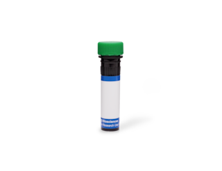
BD Transduction Laboratories™ Purified Mouse Anti-Lamin A/C
Clone 14/LaminAC (RUO)


Western blot analysis of Lamin A/C on a HeLa lysate. Lane 1: 1:1000, Lane 2: 1:2000, Lane 3: 1:4000 dilution of the anti- Lamin A/C antibody.


Regulatory Status Legend
Any use of products other than the permitted use without the express written authorization of Becton, Dickinson and Company is strictly prohibited.
Preparation And Storage
Product Notices
- Since applications vary, each investigator should titrate the reagent to obtain optimal results.
- Please refer to www.bdbiosciences.com/us/s/resources for technical protocols.
- Caution: Sodium azide yields highly toxic hydrazoic acid under acidic conditions. Dilute azide compounds in running water before discarding to avoid accumulation of potentially explosive deposits in plumbing.
- Source of all serum proteins is from USDA inspected abattoirs located in the United States.
Companion Products


The nuclear envelope (NE) is a specialized extension of the ER that contains numerous pore complexes interconnected with the nuclear lamina. The nuclear lamina composes the structural framework for the NE and serves as a chromatin anchor site, thus playing a major role in interphase nuclear organization. The major component of nuclear lamina are intermediate filament-like proteins called lamins. In mammalian somatic cells, there are three major lamins, A, B1, and C, and two minor lamins, B2 and A10. A-type lamins (A, A10, and C) are encoded by a single gene and are produced by alternative splicing, while B-type lamins (B1 and B2) are encoded by separate genes. B-type lamins are found in all nucleated somatic cells, while the expression of A-type lamins are developmentally regulated. Mice lacking lamin A show no overt abnormalities until postnatal development when perturbations in nuclear envelop structure correlate with the appearance of muscular dystrophy. In addition, lamin A is mutated in lipodystrophy, a disorder characterized by reduction in subcutaneous adipose tissue. Thus, lamin A and C may be important for nuclear envelope formation during postnatal cell differentiation.
This antibody is routinely tested by western blot analysis. Other applications were tested at BD Biosciences Pharmingen during antibody development only or reported in the literature.
Development References (3)
-
McKeon FD, Kirschner MW, Caput D. Homologies in both primary and secondary structure between nuclear envelope and intermediate filament proteins. Nature. 1986; 319(6053):463-468. (Biology). View Reference
-
Shackleton S, Lloyd DJ, Jackson SN, et al. LMNA, encoding lamin A/C, is mutated in partial lipodystrophy. Nat Genet. 2000; 24(2):153-156. (Biology). View Reference
-
Sullivan T, Escalante-Alcalde D, Bhatt H, et al. Loss of A-type lamin expression compromises nuclear envelope integrity leading to muscular dystrophy. J Cell Biol. 1999; 147(5):913-920. (Biology). View Reference
Please refer to Support Documents for Quality Certificates
Global - Refer to manufacturer's instructions for use and related User Manuals and Technical data sheets before using this products as described
Comparisons, where applicable, are made against older BD Technology, manual methods or are general performance claims. Comparisons are not made against non-BD technologies, unless otherwise noted.
For Research Use Only. Not for use in diagnostic or therapeutic procedures.