-
Your selected country is
Norway
- Change country/language
Old Browser
This page has been recently translated and is available in French now.
Looks like you're visiting us from {countryName}.
Would you like to stay on the current country site or be switched to your country?
Multiparameter flow cytometric analysis of CD4 expression on Human peripheral blood leukocyte populations. Human whole blood was stained with either BD Horizon™ BV480 Mouse IgG1, κ Isotype Control (Cat. No. 565652; Left Plot) or BD Horizon™ BV480 Mouse Anti-Human CD4 antibody (Cat. No. 569781; Right Plot) at 1 µg/test. Erythrocytes were lysed with BD Pharm Lyse™ Lysing Buffer (Cat. No. 555899). The bivariate pseudocolor density plot showing the correlated expression of CD4 (or Ig Isotype control staining) versus side-light scatter signals (SSC-A) was derived from gated events with the forward and side light-scatter characteristics of viable leukocytes. Flow cytometry and data analysis were performed using a BD LSRFortessa™ X-20 Cell Analyzer and FlowJo™ Software. Data shown on this Technical Data Sheet are not lot specific.
BD Horizon™ BV480 Mouse Anti-Human CD4
Regulatory Status Legend
Any use of products other than the permitted use without the express written authorization of Becton, Dickinson and Company is strictly prohibited.
Preparation And Storage
Recommended Assay Procedures
BD® CompBeads can be used as surrogates to assess fluorescence spillover (compensation). When fluorochrome conjugated antibodies are bound to BD® CompBeads, they have spectral properties very similar to cells. However, for some fluorochromes there can be small differences in spectral emissions compared to cells, resulting in spillover values that differ when compared to biological controls. It is strongly recommended that when using a reagent for the first time, users compare the spillover on cells and BD® CompBeads to ensure that BD® CompBeads are appropriate for your specific cellular application.
For optimal and reproducible results, BD Horizon Brilliant™ Stain Buffer should be used anytime BD Horizon Brilliant dyes are used in a multicolor flow cytometry panel. Fluorescent dye interactions may cause staining artifacts which may affect data interpretation. The BD Horizon Brilliant Stain Buffer was designed to minimize these interactions. When BD Horizon Brilliant Stain Buffer is used in the multicolor panel, it should also be used in the corresponding compensation controls for all dyes to achieve the most accurate compensation. For the most accurate compensation, compensation controls created with either cells or beads should be exposed to BD Horizon Brilliant Stain Buffer for the same length of time as the corresponding multicolor panel. More information can be found in the Technical Data Sheet of the BD Horizon Brilliant Stain Buffer (Cat. No. 563794/566349) or the BD Horizon Brilliant Stain Buffer Plus (Cat. No. 566385).
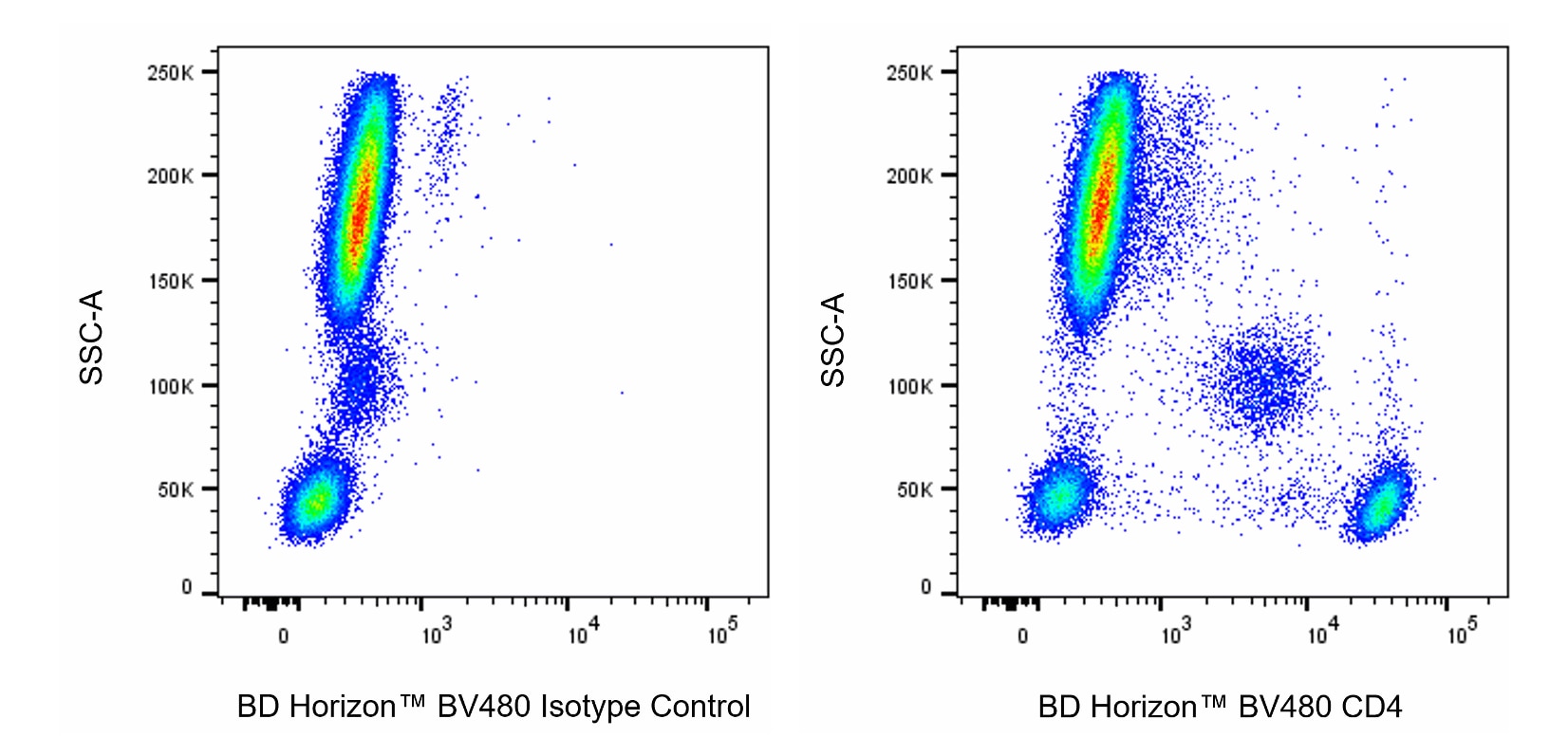
For Immunofluorescence Applications:
The use of a mounting reagent (eg, ProLong® Gold) is highly recommended to maximize the photostability of BV480. For confocal microscopy systems, a 440 nm laser is the optimal excitation source and the recommended emission filter is a 485/20 nm bandpass filter.
For epifluorescence microscopes with broad spectrum excitation sources, the recommended excitation and emission filters are 445/20 nm and 485/20 nm bandpass filters, respectively. For specific multicolor imaging applications, the exact filter configurations should be optimized by the end user. For additional instrument/filter configuration information, please visit http://www.bdbiosciences.com/research/cellularimaging.
Product Notices
- Please refer to www.bdbiosciences.com/us/s/resources for technical protocols.
- Since applications vary, each investigator should titrate the reagent to obtain optimal results.
- An isotype control should be used at the same concentration as the antibody of interest.
- Caution: Sodium azide yields highly toxic hydrazoic acid under acidic conditions. Dilute azide compounds in running water before discarding to avoid accumulation of potentially explosive deposits in plumbing.
- For fluorochrome spectra and suitable instrument settings, please refer to our Multicolor Flow Cytometry web page at www.bdbiosciences.com/colors.
- Human donor specific background has been observed in relation to the presence of anti-polyethylene glycol (PEG) antibodies, developed as a result of certain vaccines containing PEG, including some COVID-19 vaccines. We recommend use of BD Horizon Brilliant™ Stain Buffer in your experiments to help mitigate potential background. For more information visit https://www.bdbiosciences.com/en-us/support/product-notices.
- Please refer to http://regdocs.bd.com to access safety data sheets (SDS).
- For U.S. patents that may apply, see bd.com/patents.
Companion Products
The RPA-T4 monoclonal antibody specifically binds to CD4, a 59 kDa single-chain transmembrane glycoprotein that is expressed on T-helper/inducer cell populations. CD4 is also expressed on thymocyte subsets and at lower levels on monocytes and macrophages. CD4 functions as a co-receptor in MHC class II-restricted antigen-induced T cell activation and as a receptor for human immunodeficiency viruses (HIV). This antibody binds to the D1 domain (CDR1 and CDR3 epitopes) of the CD4 antigen and reacts with approximately 80% of thymocytes and 45% of peripheral blood lymphocytes. RPA-T4 is capable of blocking HIV-1, gp120, and inhibits syncytium formation.
Development References (6)
-
Ho TH, Pfeffer K, Weiss GJ, Ruiz Y, Lake DF. Identification of a CD4+ T cell line with Treg-like activity.. Hum Immunol. 2022; 83(4):281-294. (Clone-specific: Flow cytometry). View Reference
-
Knapp W. W. Knapp .. et al., ed. Leucocyte typing IV : white cell differentiation antigens. Oxford New York: Oxford University Press; 1989:1-1182.
-
Moebius U. Cluster report: CD4. In: Knapp W. W. Knapp .. et al., ed. Leucocyte typing IV : white cell differentiation antigens. Oxford New York: Oxford University Press; 1989:314-330.
-
Piatier-Tonneau D. CD4 workshop panel report. In: Kishimoto T. Tadamitsu Kishimoto .. et al., ed. Leucocyte typing VI : white cell differentiation antigens : proceedings of the sixth international workshop and conference held in Kobe, Japan, 10-14 November 1996. New York: Garland Pub.; 1997:49-54.
-
Pucino V, Certo M, Bulusu V, et al. Lactate Buildup at the Site of Chronic Inflammation Promotes Disease by Inducing CD4+ T Cell Metabolic Rewiring.. Cell Metab. 2019; 30(6):1055-1074.e8. (Clone-specific: Flow cytometry). View Reference
-
Schlossman SF. Stuart F. Schlossman .. et al., ed. Leucocyte typing V : white cell differentiation antigens : proceedings of the fifth international workshop and conference held in Boston, USA, 3-7 November, 1993. Oxford: Oxford University Press; 1995.
Please refer to Support Documents for Quality Certificates
Global - Refer to manufacturer's instructions for use and related User Manuals and Technical data sheets before using this products as described
Comparisons, where applicable, are made against older BD Technology, manual methods or are general performance claims. Comparisons are not made against non-BD technologies, unless otherwise noted.
For Research Use Only. Not for use in diagnostic or therapeutic procedures.