Old Browser
This page has been recently translated and is available in French now.
Looks like you're visiting us from {countryName}.
Would you like to stay on the current country site or be switched to your country?
BD Pharmingen™ Propidium Iodide Staining Solution
(RUO)


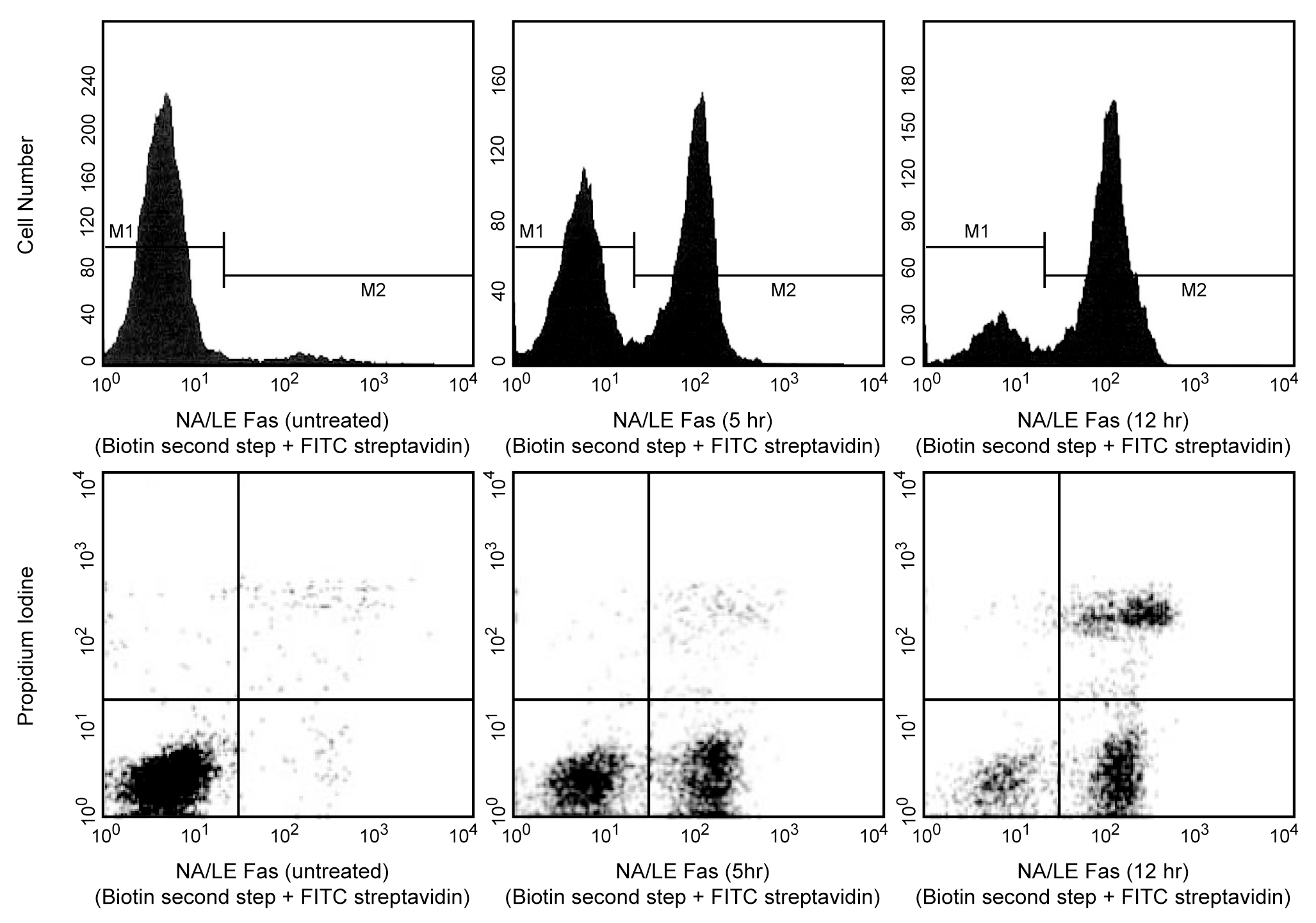
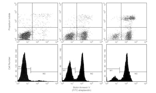
Flow cytometric analysis of cells following induction of apoptosis. Jurkat leukemia cells were left untreated (left top & left bottom panels) or treated for 5 hours (middle top & middle bottom panels) or 12 hours (right top & right bottom panels) with anti-human Fas antibody (clone DX2, Cat. No. 555670) and Protein G (the addition of Protein G enhances the ability of DX2 to induce apoptosis, presumably by cross-linking Fas). Cells were incubated with Annexin V-Biotin, (Cat. No. 556417) followed by incubation with SAv-FITC (Cat. No. 554060) in Propidium Iodide (PI) Staining Solution (Cat. No. 556463). Cells were then analyzed by flow cytometry. Untreated cells were primarily Annexin V-Biotin and PI negative, indicating that they were viable and not undergoing apoptosis. After a 5 hour treatment with DX2, there were two populations of cells: Cells undergoing apoptosis (Annexin V-Biotin positive and PI negative), and cells that were viable and not undergoing apoptosis (Annexin V-Biotin and PI negative). After a 12 hour treatment with DX2, three populations of cells were identified: Cells that had already died or were in late stage of apoptosis (Annexin V-Biotin and PI positive), cells undergoing apoptosis (Annexin V-Biotin positive and PI negative), and cells that were viable and not undergoing apoptosis (Annexin V-Biotin and PI negative).

BD Pharmingen™ Propidium Iodide Staining Solution
Regulatory Status Legend
Any use of products other than the permitted use without the express written authorization of Becton, Dickinson and Company is strictly prohibited.
Description
The Prodidium Iodide (PI) Staining Solution may be used to assess plasma membrane (PM) integrity in Annexin V apoptosis assays. PI is a fluorescent vital dye that stains DNA. It does not cross the PM of cells that are viable or in the early stages of apoptosis because they maintain PM integrity. In contrast those cells in the late stages of apoptosis or already dead have lost PM integrity and are permeable to PI. PI is detected in the orange range of the spectrum using a 562-588 nm band pass filter. Annexin V binds to cells early in apoptosis, and continues to be bound through cell death. PI is used in two-color Annexin V flow cytometric assays to distinguish cells that are in the earlier stages of apoptosis (Annexin V positive, PI negative) from those that are in the later stages of apoptosis or already dead (Annexin V positive, PI positive).
Preparation And Storage
The PI Staining Solution is composed of 50 µg PI/ml in PBS (pH 7.4) and is 0.2 µm sterile filtered.
Recommended Assay Procedures
Recommended Assay Procedure:
The PI Staining Solution is designed for use in two-color Annexin V flow cytometric assays. It can be used in conjunction with Annexin V conjugated to fluorescein isothiocyanate (Annexin V-FITC, Cat. No. 556419) or biotinylated Annexin V (Annexin V-Biotin, Cat. No. 556417). Suggested amounts to use are 10 µl/test (1x10e6 cells).
Caution: Propidium Iodide is a potential carcinogen. Avoid contact with skin and eyes. Wear suitable protective clothing, gloves, and eye/face protection.
Product Notices
- Since applications vary, each investigator should titrate the reagent to obtain optimal results.
- This reagent has been pre-diluted for use at the recommended Volume per Test. We typically use 1 × 10^6 cells in a 100-µl experimental sample (a test).
- Please refer to www.bdbiosciences.com/us/s/resources for technical protocols.
Companion Products


.png?imwidth=320)

Development References (1)
-
Martin SJ, Reutelingsperger CP, McGahon AJ, et al. Early redistribution of plasma membrane phosphatidylserine is a general feature of apoptosis regardless of the initiating stimulus: inhibition by overexpression of Bcl-2 and Abl. J Exp Med. 1995; 182(5):1545-1556. (Methodology: Apoptosis, Flow cytometry). View Reference
Please refer to Support Documents for Quality Certificates
Global - Refer to manufacturer's instructions for use and related User Manuals and Technical data sheets before using this products as described
Comparisons, where applicable, are made against older BD Technology, manual methods or are general performance claims. Comparisons are not made against non-BD technologies, unless otherwise noted.
For Research Use Only. Not for use in diagnostic or therapeutic procedures.
Report a Site Issue
This form is intended to help us improve our website experience. For other support, please visit our Contact Us page.